Session Information
Date: Sunday, October 7, 2018
Session Title: Huntington's Disease
Session Time: 1:45pm-3:15pm
Location: Hall 3FG
Objective: The aim of our study is to characterize in detail the white matter (WM) microstructure alterations in Huntington Disease (HD) using the diffusion tensor imaging (DTI) analysis on magnetic resonance imaging (MRI).
Background: Imaging studies in patients with HD may help to determine differential vulnerability of CNS structures to the neurodegenerative process as well as to identify precisely when neurodegeneration starts, how it progresses and the associated triggers [1].
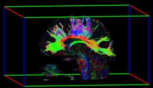
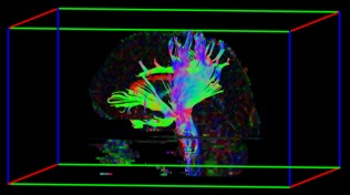
Methods: We obtained DTI (32 directions) acquired at 3.0T from 37 healthy volunteers and 36 patients (genetically confirmed), balanced for age (p=0.9) and gender (p=0.9). Patients underwent neurological (Unified Huntington’s disease rating scale – UHDRS) and cognitive (Montreal cognitive assessment – MOCA) evaluations. Diffusion Tensor Images were processed with ExploreDTI/MATLAB-2014 (www.exploredti.com). Ten tracts [3 parts of the corpus callosum (CC), Corticospinal tract (CST), Inferior Fronto Occipital (IFO) tract, Inferior Longitudinal Fasciculus (ILF), dorsal and parahippocampal cingulum (PH-CINGULUM), uncinate and body of fornix (FORNIX)] were delineated by semi-automatic deterministic tractography to yield fractional anisotropy (FA) (Figures 1 and 2). SPSS22 was used for correlations, univariate, multivariate analyses and Chi-square test.
Results: Multivariate analyses with Repeated-measures ANOVA for bilateral tracts revealed significant FA reduction mainly on cingulum and PH-cingulum (p<0.004, with Bonferroni correction). MANOVA of Corpus Callosum segments and FORNIX showed reduced FA values (p<0.0125 with Bonferroni correction) in patients with HD. While there was no significant correlation between FA values and CAG repeat expansion, we identified a significant correlation between UHDRS and CST (left r=-0.575, right r=-0.45, p<0.005), IFO (left r=-0.51, left r=-0.58, p<0.002) and left PH-CINGULUM (r=-0.48, p=0.003). In addition, there was a positive correlation between MOCA and FA values in PH-CINGULUM (left r=0.54, right r=0.59, p<0.002).
Conclusions: WM microstructural alterations were widespread, affecting midline and bilateral structures in patients with HD [2]. The abnormal tracts are responsible for sensorimotor integration, motor control and planning, visuospatial function and emotional processing. Prospective studies are underway in order to characterize how the pattern of WM alterations progresses over time.
References: [1] Wu D, et al. Mapping the order and pattern of brain structural MRI changes using change-point analysis in premanifest Huntington’s disease. PREDICT-HD Investigators and Coordinators of the Huntington Study Group. Hum Brain Mapp. 2017 Oct;38(10):5035-5050. [2] Saba RA, et al. Diffusion tensor imaging of brain white matter in Huntington gene mutation individuals. Arq Neuropsiquiatr. 2017 Aug;75(8):503-508.
To cite this abstract in AMA style:
P. Azevedo, L. Piovesana, M. Nogueira, R. Guimarães, A. Amato Filho, F. Cendes, Í. Lopes-Cendes, C. Yasuda. White matter microstructural alterations in Huntington Disease: When neurodegeneration starts? [abstract]. Mov Disord. 2018; 33 (suppl 2). https://www.mdsabstracts.org/abstract/white-matter-microstructural-alterations-in-huntington-disease-when-neurodegeneration-starts/. Accessed December 13, 2025.« Back to 2018 International Congress
MDS Abstracts - https://www.mdsabstracts.org/abstract/white-matter-microstructural-alterations-in-huntington-disease-when-neurodegeneration-starts/


