Session Information
Date: Monday, September 23, 2019
Session Title: Huntington’s Disease
Session Time: 1:45pm-3:15pm
Location: Agora 3 West, Level 3
Objective: Our study investigated the impact of Huntington disease (HD) on brain connectivity, through resting-state (RS) fMRI, among the most critical regions associated with physiopathology.
Background: HD is an autosomal dominant neurodegenerative disorder that progressively affects motor, cognitive and behavioral control. Lately, there has been an effort to detail the cerebral dysfunction in HD patients [1]. Here we used resting-state fMRI to investigate alterations in functional connectivity.
Method: Resting-state fMRI (RS-fMRI) was acquired on 3T-PHILIPS from 22 patients (15 women, median age 52 years, range 25-68) and 22 controls (15 women, median 51.5 years, range 24-64), matched by age (p=0.9) and sex (p=1). RS images were preprocessed with UF2C-toolbox (running on MATLAB2014/SPM12). The same software was used for ROI parcellation, extraction of time series, matrix construction, and statistical analysis. In the primary analysis, we selected 50 anatomical regions of interest (ROI) to investigate intranetwork and internetwork connectivity. The secondary analysis involved functional 18 ROIs selected from 4 RS-networks (Basal ganglia, sensorimotor, L/R Executive control networks). Reported results have a 0.05 alpha (FDR corrected for multiple comparisons).
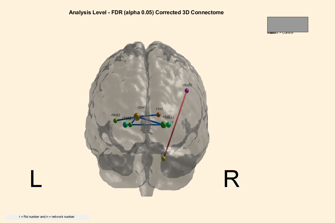
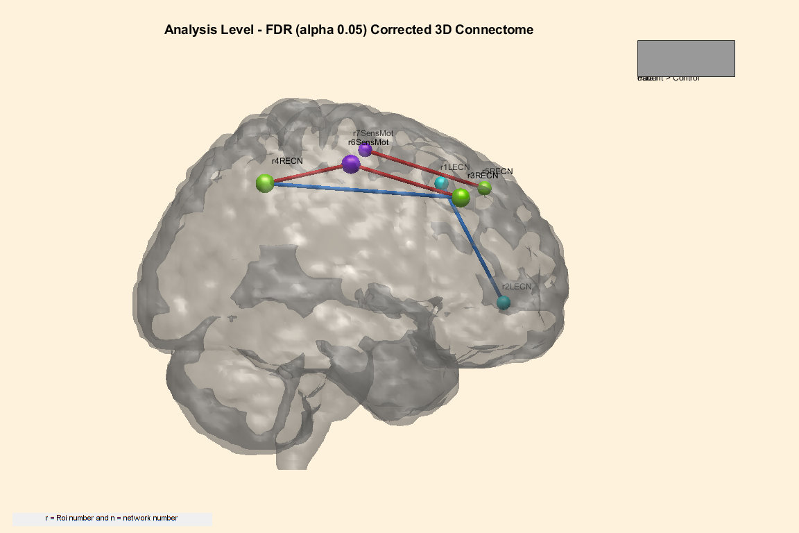
Results: Compared to controls, there were intranetwork and internetwork dysfunctions. The patient group presented reduced connectivity compared to controls between the following regions (p=0.005) [Figure 1]: right (R) and left (L) caudate; L-caudate with R/L pallidum and L-frontal operculum; R-pallidum with R/L putamen. Interestingly, there was only one increased connectivity measure in the HD subjects between the anatomical regions of R-cerebellum and R-angular gyrus. Concerning the functional networks [Figure 2], HD patients presented increased connectivity between R-executive control network and sensorimotor area (p=0.038).
Conclusion: These results suggest that HD individuals present abnormal interactions of large-scale brain network (mostly reduced connectivity). Although all patients were right-handed, network dysfunction was similar in both hemispheres. The unique increased connection in patients involved an association area, probably associated with reading ability and mathematical processing in the dominant hemisphere [2]. Further investigation is necessary to evaluate the impact of such dysfunctional networks on both cognition, behavior and motor alterations.
References: [1]Mapping the order and pattern of brain structural MRI changes using change-point analysis in premanifest Huntington’s disease; [2] ww.atlasbrain.com/enx/glossary/A_glossary.htm.
To cite this abstract in AMA style:
P. de Azevedo, M. Nogueira, L. Ribeiro, B. de Campos, R. Guimarães, L. Piovesana, F. Cendes, I. Lopes-Cendes, C. Yasuda. What connectivity tells us about neurodegeneration in Huntington disease? A fMRI study [abstract]. Mov Disord. 2019; 34 (suppl 2). https://www.mdsabstracts.org/abstract/what-connectivity-tells-us-about-neurodegeneration-in-huntington-disease-a-fmri-study/. Accessed April 18, 2025.« Back to 2019 International Congress
MDS Abstracts - https://www.mdsabstracts.org/abstract/what-connectivity-tells-us-about-neurodegeneration-in-huntington-disease-a-fmri-study/


