Category: Parkinson's Disease: Neuroimaging
Objective: Our study aimed to analyze the alterations in visual network functional connectivity (FC) in subjects with PD and concomitant visual disturbance (VD) and/or visual hallucination (VH) compared to PD subjects with no visual disorder.
Background: Parkinson’s disease (PD) is frequently accompanied by visual disturbances. The spectrum of visual disturbances arises from blurred vision and can get as serious as visual hallucinations. The mechanism behind this spectrum of disturbances has not been cleared yet.
Method: 18 PD subjects with concomitant VD and/or VH and 25 PD subjects with no visual disorders (NVD group) were enrolled in this study. 9 out of 18 subjects had only VD (VD group) and the other 9 experienced VH ± VD (VHD group). Resting-state fMRI scan was performed for subjects using a 3T Siemens scanner with 64 channels head and neck coil. Seed-to-voxel analysis was performed using Conn toolbox with four visual networks including the visual medial, visual occipital, visual left lateral, and visual right lateral as seed regions. Group-based analysis was then performed between the three groups. A voxel threshold p-value set of <0.01 was used, with the results corrected for multiple comparisons using fdr with a significance level of 0.05. Age, gender, and disease severity based on the Unified Parkinson’s Disease Rating Scale (UPDRS III) were used as confounding variables.
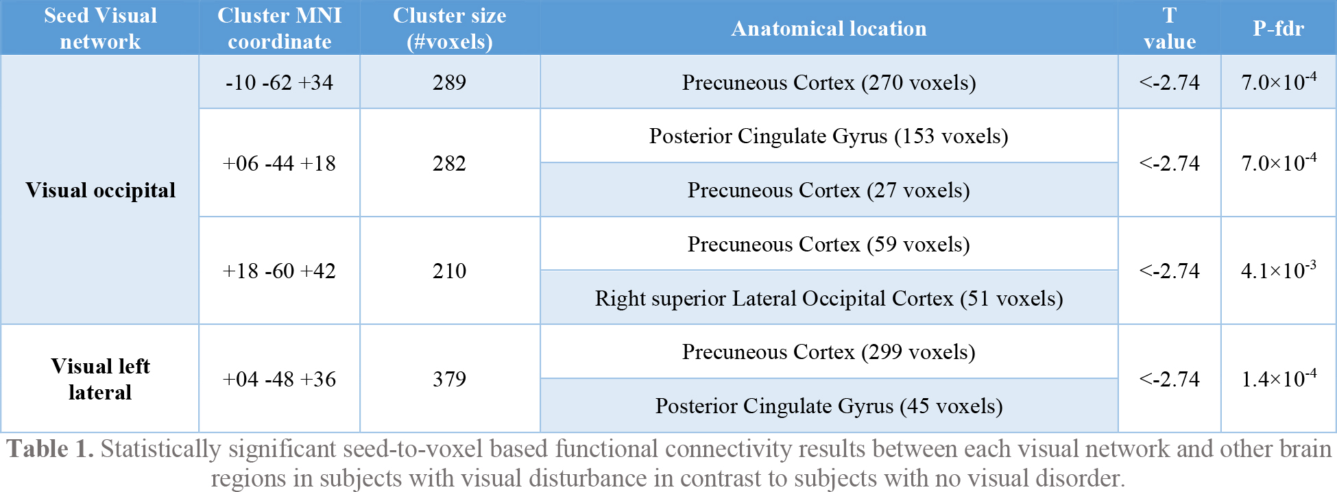
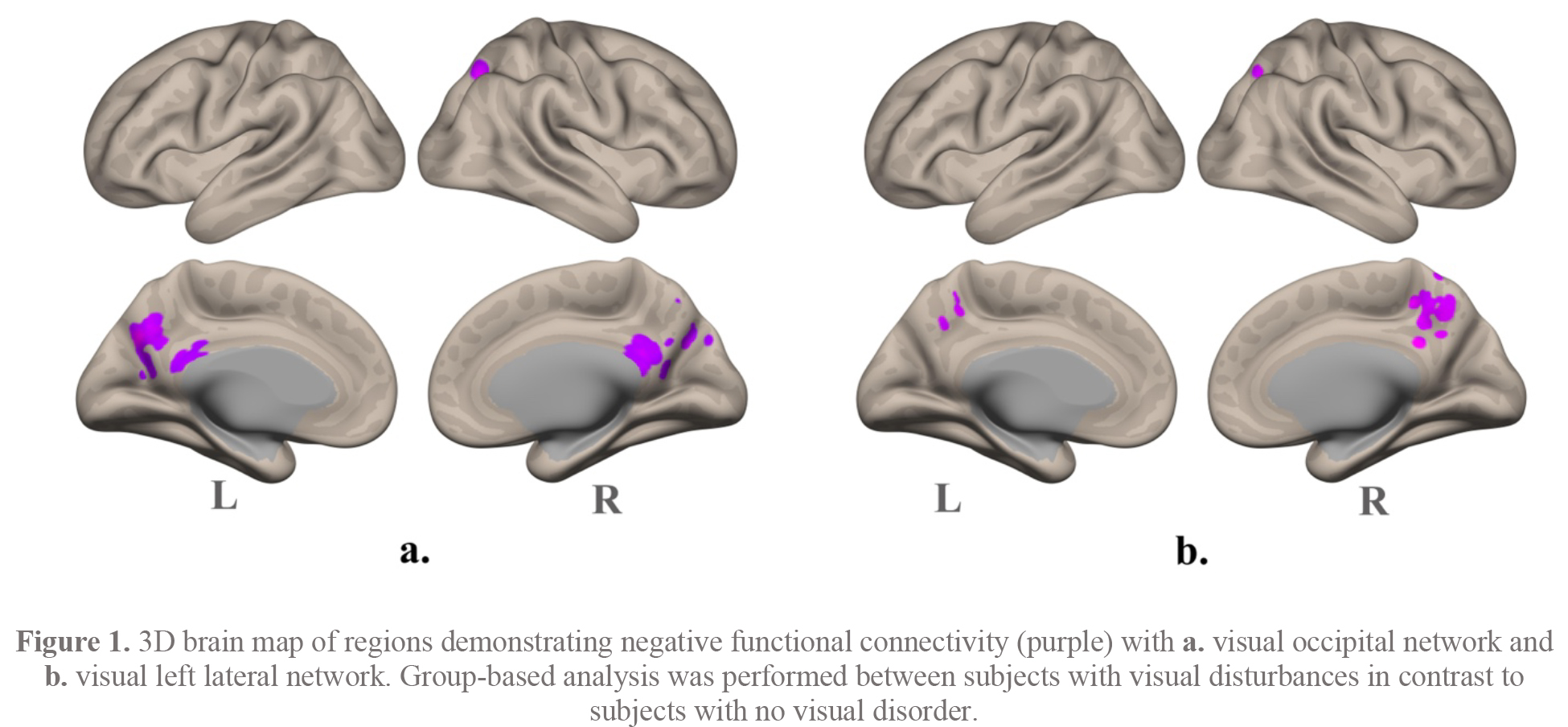
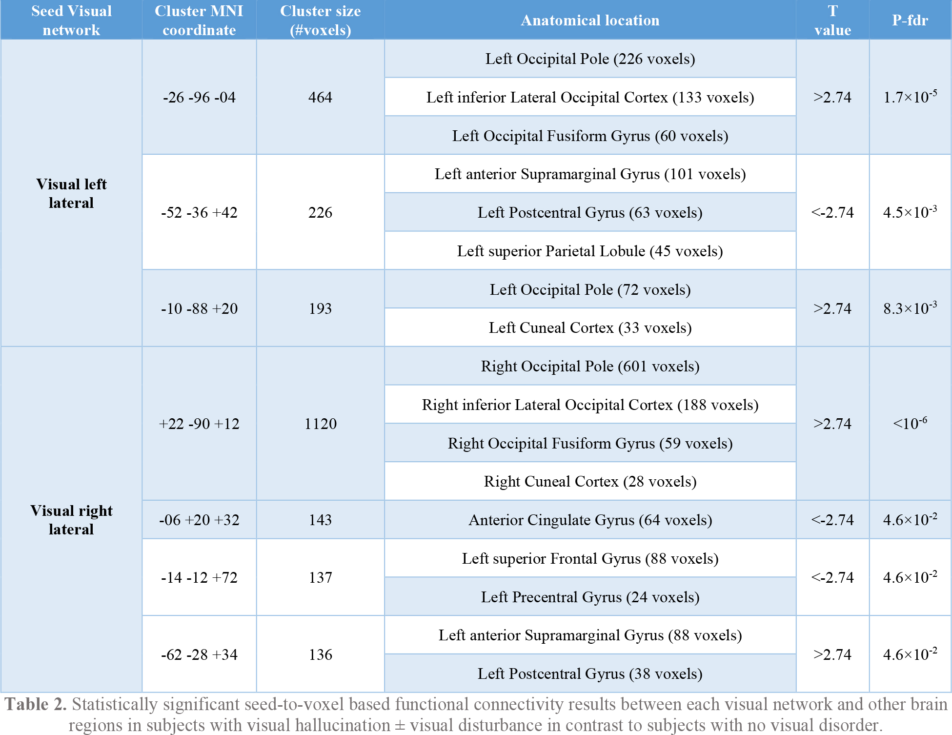
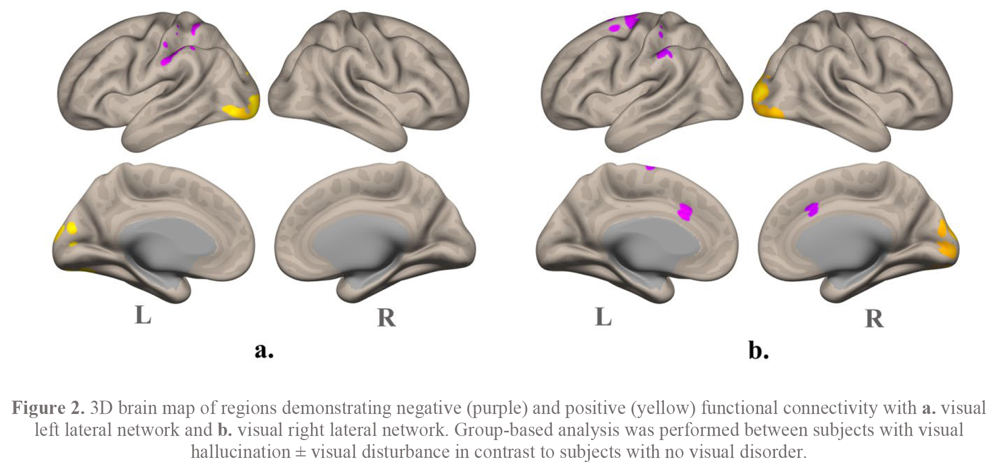
Results: The VD group compared to the NVD group showed decreased FC between occipital and left lateral visual networks and other brain regions. The VHD group compared to the NVD group showed altered FC between the right and left lateral networks and other brain regions. Finally, analysis of the VHD group compared to the VD group showed increased FC between medial and right lateral networks and other brain regions. Each table demonstrates clusters’ alteration details for each group analysis. Figures represent the size and location of each group analysis on the brain.
Conclusion: Altered FC was observed between visual networks and other brain regions involved in integrating information, perception of the environment, and reactivity in PD patients with concomitant VH and/or VD compared to patients with no visual disorders. Visual network alterations have been seen in psychiatric patients with hallucinations. Understanding the nature of changes in PD can lead to a better insight into the pathophysiology of VD and/or VH.
References: 1. Kesler A, Korczyn AD. Visual disturbances in Parkinson’s disease. Practical Neurology. 2006;6(1):28-33.
2. Barnes J. Visual hallucinations in Parkinson’s disease: a review and phenomenological survey. Journal of Neurology, Neurosurgery & Psychiatry. 2001;70(6):727-33.
3. Goetz CG, Tilley BC, Shaftman SR, Stebbins GT, Fahn S, Martinez-Martin P, et al. Movement Disorder Society-sponsored revision of the Unified Parkinson’s Disease Rating Scale (MDS-UPDRS): Scale presentation and clinimetric testing results. Movement Disorders. 2008;23(15):2129-70.
To cite this abstract in AMA style:
O. Shoraka, M. Syed, I. Shelley, K. Shivock, TW. Liang, RC. Sergott, I. Fayed, A. Sharan, M. Alizadeh. Visual networks connectivity in subjects with Parkinson’s disease and visual disturbances using resting state fMRI [abstract]. Mov Disord. 2023; 38 (suppl 1). https://www.mdsabstracts.org/abstract/visual-networks-connectivity-in-subjects-with-parkinsons-disease-and-visual-disturbances-using-resting-state-fmri/. Accessed February 23, 2026.« Back to 2023 International Congress
MDS Abstracts - https://www.mdsabstracts.org/abstract/visual-networks-connectivity-in-subjects-with-parkinsons-disease-and-visual-disturbances-using-resting-state-fmri/


