Category: Parkinson's Disease: Cognitive functions
Objective: Our aim is to understand MRT abnormalities in PD using fMRI and whether it can be used as a clinical marker for disease progression in Early Onset Parkinson’s Disease (EOPD).
Background: EOPD is defined by age of onset between 40 and 60 and has no known genetic etiology. PD initially presents with unilateral onset of motor deficits and progresses to bilaterality. Hemisphere-specific cognitive and behavioral abnormalities are proposed to be related to side of disease onset. Minimal cognitive impairment may go undetected as routine assessments lack sensitivity to recognize subtle dysfunction in EOPD patients. One such measure being considered for assessing early visuospatial deficits in PD is the MRT. EOPD patients with left-sided onset motor symptoms (LPD) should have greater right hemispherical dysfunction and vice versa. As most visuospatial information is processed by the right posterior parietal lobe, LPD patients may have significant visuospatial deficits compared to right onset PD patients (RPD).
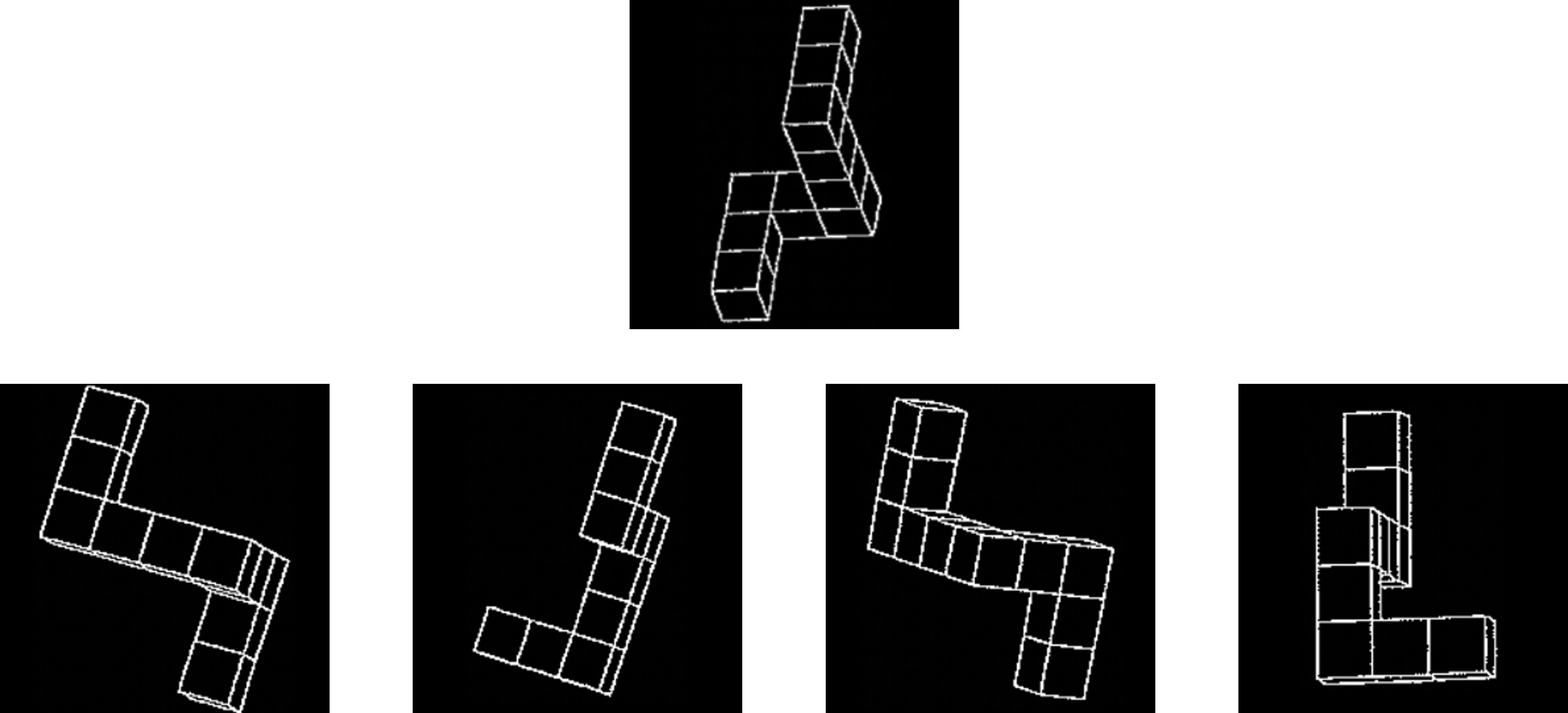
Method: MRT (example in figure 1) scores were analyzed for 26 Stage 1 RPD subjects, 20 LPD subjects, 19 EOPD subjects with bilateral symptoms and 14 non-PD controls. Structural MRIs were used to obtain volumetric data for parietal lobe regions and their correlation to MRT scores. We also plan to use fMRI blood oxygen level-dependent (BOLD) imaging to understand the mechanistic basis of brain activation differences between EOPD subjects and controls during MRT in ongoing studies.
Results: MRT scores in the LPD stage 1 subjects (p= 0.024) and bilateral (p=0.034) subjects were significantly lower than RPD subjects. However, MRT scores in the LPD stage 1 subjects were not statistically significantly different than stage subjects (p=0.690). Structural MRI in 17 RPD and 14 LPD subjects showed left (p<0.012) and right (p<0.0017) inferior parietal lobe volumes differed between males and approached significance in females. MRT scores positively correlated with left Inferior parietal lobe volume in LPD male and female subjects combined (r=0.71, p<0.031).
Conclusion: The findings suggest that MRT is a sensitive test to detect hemisphere specific visuospatial differences in PD patients. Combined with data from fMRI imaging this could be utilized as an early indicator of cognitive decline and provide possible targets for delaying disease progression in EOPD.
To cite this abstract in AMA style:
J. Razzaque, B. Mullen, C. Dougherty, A. Cotton, D. Wagner, S. Ravi, K. Le, K. Iyer, M. Subramanian, J. Wang, K. Venkiteswaran, T. Subramanian, P. Eslinger. Understanding the Mechanisms of Brain Dysfunction when Performing the Mental Rotation Task (MRT) in Stage 1 and Stage 2 EOPD using brain imaging [abstract]. Mov Disord. 2023; 38 (suppl 1). https://www.mdsabstracts.org/abstract/understanding-the-mechanisms-of-brain-dysfunction-when-performing-the-mental-rotation-task-mrt-in-stage-1-and-stage-2-eopd-using-brain-imaging/. Accessed April 20, 2025.« Back to 2023 International Congress
MDS Abstracts - https://www.mdsabstracts.org/abstract/understanding-the-mechanisms-of-brain-dysfunction-when-performing-the-mental-rotation-task-mrt-in-stage-1-and-stage-2-eopd-using-brain-imaging/

