Session Information
Date: Wednesday, September 25, 2019
Session Title: Surgical Therapy
Session Time: 1:15pm-2:45pm
Location: Les Muses Terrace, Level 3
Objective: To report on radiological and topographical features of the lesion observed at 24-hours and 3-months following Transcranial MR guided FUS (tcMRgFUS) thalamotomy in a cohort of medically refractory essential tremor patients, and to correlate them with the procedure profiles and the clinical outcomes.
Background: Unilateral tcMRgFUS thalamotomy has emerged as a safe and effective alternative for the treatment of medically-refractory essential tremor [1]. Thalamic lesions following tcMRgFUS have been characterized as a set of three concentric zones with different tissue properties. On T2w images, inner zones 1 and 2, i.e. lesion core and periphery, represent coagulation necrosis and cytotoxic edema, and the larger and fuzzier outer ring (zone 3) vasogenic edema [2].
Method: Forty patients with medication-refractory essential tremor were treated with unilateral tcMRgFUS thalamotomy. Clinical and radiological data were prospectively acquired at baseline, immediately and at 3-months after treatment. Treatment efficacy was assessed with Clinical Rating Scale for Tremor (CRST). Lesions were manually segmented on T1, T2 and susceptibility weighted images, and three-dimensional topographical analysis was then carried out. Statistical comparisons were performed using nonparametric statistics.
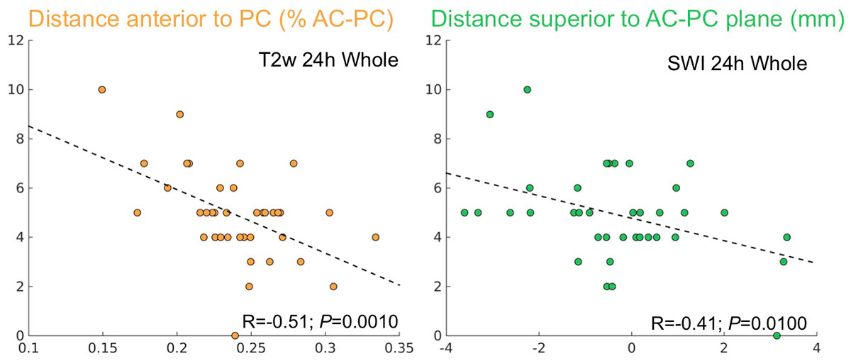
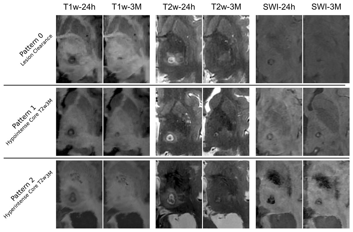
Results: The major clinical improvement was correlated with a more ventrocaudal lesion (P<0.05) [Figure 1], greater lesion volume (P<0.05), whereas the largest lesions accounted for the occurrence of gait imbalance (P<0.05). The volume of the lesion was significantly predicted (P<0.05) by the number of sonications surpassing 52ºC. Furthermore, three different lesion patterns were identified on T2w images at 3-months [Figure 2], which depended on age and the treatment profile but had no relation with the clinical outcome.
Conclusion: We provide a comprehensive characterization of the thalamic tcMRgFUS lesion including radiological and topographical analysis. Three-dimensional topographical analysis revealed a significant association between the location and volume of the lesion and the clinical outcome, although largest lesions were also related to the presence of adverse events. The number of moderate temperature sonications correlated with the final lesion volumes thus empowering the importance of the sonications below 55ºC.
References: 1- Elias W.J., Lipsman N., Ondo W.G., Ghanouni P., Kim, Y.G., Lee W., et al: A Randomized Trial of Focused Ultrasound Thalamotomy for Essential Tremor. N. Engl. J. Med. 375, 730–739, 2016. 2- Wintermark M, Druzgal J, Huss DS, Khaled M a, Monteith S, Raghavan P, et al: Imaging Findings in MR Imaging–Guided Focused Ultrasound Treatment for Patients with Essential Tremor. Am J Neuroradiol AJNR 35:891–96, 2014.
To cite this abstract in AMA style:
D. Urso, J. Pineda-Pardo, R. Martínez-Fernández, R. Rodríguez-Rojas, M. Del-Alamo, P. Millar-Vernetti, JU. Mañez, F. F. Hernández-Fernández, E. de Luis-Pastor, L. Vela, JA. Obeso. Transcranial MR guided FUS thalamotomy in essential tremor: a comprehensive lesion characterization [abstract]. Mov Disord. 2019; 34 (suppl 2). https://www.mdsabstracts.org/abstract/transcranial-mr-guided-fus-thalamotomy-in-essential-tremor-a-comprehensive-lesion-characterization/. Accessed December 27, 2025.« Back to 2019 International Congress
MDS Abstracts - https://www.mdsabstracts.org/abstract/transcranial-mr-guided-fus-thalamotomy-in-essential-tremor-a-comprehensive-lesion-characterization/


