Category: Parkinson's Disease: Neuroimaging
Objective: To assess diffusion tensor imaging differences between PD patients and healthy controls in 8 corticospinal tract segments.
Background: Parkinson’s disease (PD), despite having motor and non-motor symptoms, is mostly known by its motor features. The corticospinal tract is the major motor tract and is related to a variety of movements, such as gait, writing, typing, buttoning, which are all impaired in PD.
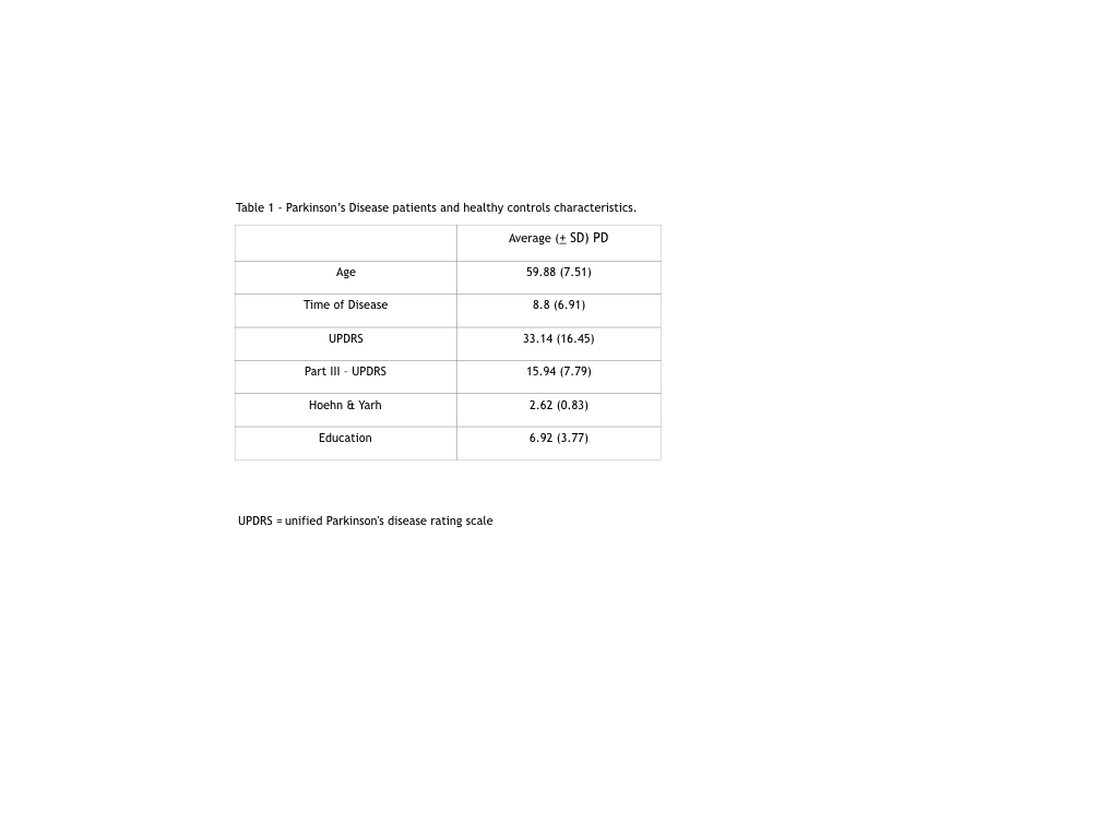
Method: We included 40 PD patients (mean age 60 years) and 129 healthy controls (mean age 58 years). Demographics are described in table 1.
DTI microstructural parameters of the cortical-spinal tract (CST) were obtained using the software ExploreDTI. The DTI images were corrected for signal drift, Gibbs’ rings, susceptibility artifact, Eddy currents, and movement and then registered to a subject T1 weighted image. We used a semi-automatic methodology for fiber tracking based on strategies drawn on a reference template. The tracts were uniformly resampled and divided into 8 segments [Figure1]. We ran a repeated measures analysis of variance with mixed designs, including the effect of segments (within-subjects), an effect of group (between-subjects) and a segment* group interaction. Significant interactions were evaluated using analysis of simple effects. We report Huynh-Feldt corrected F tests to account for deviations of sphericity assumption. Multiple comparisons were corrected using the false discovery rate (group effect) and Sidak tests (segments).
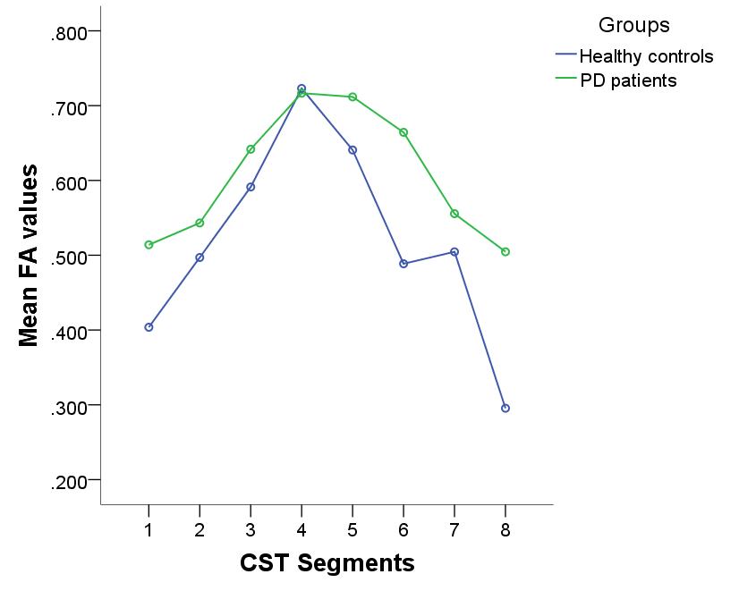
Results: We found a significant difference between PD and controls F1,161 = 48.4, p<0.001, a significant difference within segments F3,521 = 159.6, p<0.001 and significant group*segment interaction F3,521 = 18.1, p<0.001 (Figure 1). Overall, PD patients showed higher FA than controls in all (p<0.02) but the 4th segment (p=0.65).
Conclusion: The fractional anisotropy is higher in PD patients when compared to controls. Its values are also different among segments, and the same pattern is seen in controls.
To cite this abstract in AMA style:
R. Guimarães, L. Ramalho, B. Campos, L. Piovesana, P. Azevedo, F. Cendes. Tractography of the corticospinal tract in Parkinson’s disease. How does diffusion values vary along tract segments? [abstract]. Mov Disord. 2020; 35 (suppl 1). https://www.mdsabstracts.org/abstract/tractography-of-the-corticospinal-tract-in-parkinsons-disease-how-does-diffusion-values-vary-along-tract-segments/. Accessed December 31, 2025.« Back to MDS Virtual Congress 2020
MDS Abstracts - https://www.mdsabstracts.org/abstract/tractography-of-the-corticospinal-tract-in-parkinsons-disease-how-does-diffusion-values-vary-along-tract-segments/


