Session Information
Date: Tuesday, September 24, 2019
Session Title: Parkinsonisms and Parkinson-Plus
Session Time: 1:45pm-3:15pm
Location: Agora 3 West, Level 3
Objective: We used Gait-EEG to simultaneously record the three-axis acceleration and EEG during a PD patient’s timed-up-and-go (TUG) task, aimed to accurate identify and detect the freezing of gait (FOG) episodes and investigate the dynamic EEG changes in real time associated with freezing.
Background: FOG is a common gait deficit in PD patients, FOG episodes may lead to fall and reduce patients’ quality of life. Ambulatory EEG is able to track the underlying physiological changes throughout the brain in real time during turning, which is known to be the most provocative trigger for FOG. However, to analyze the EEG changes for FOG, it is necessary to accurately detect the exact onset time and ending time of FOG during walking task.
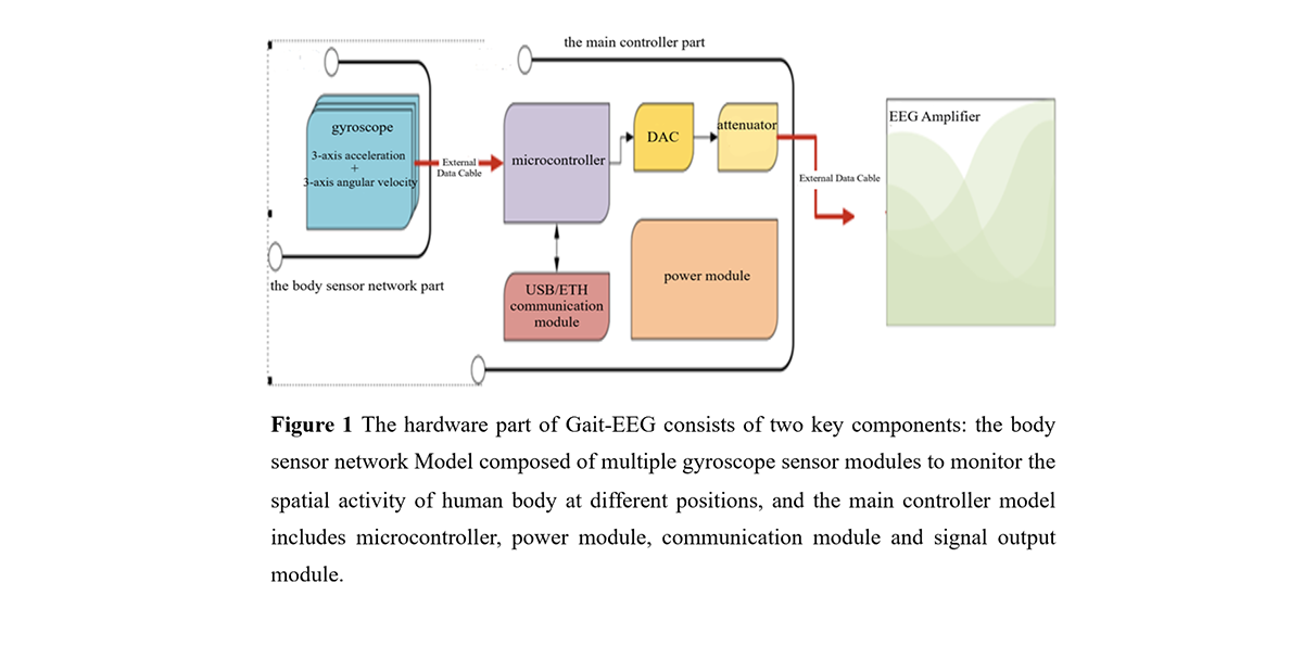
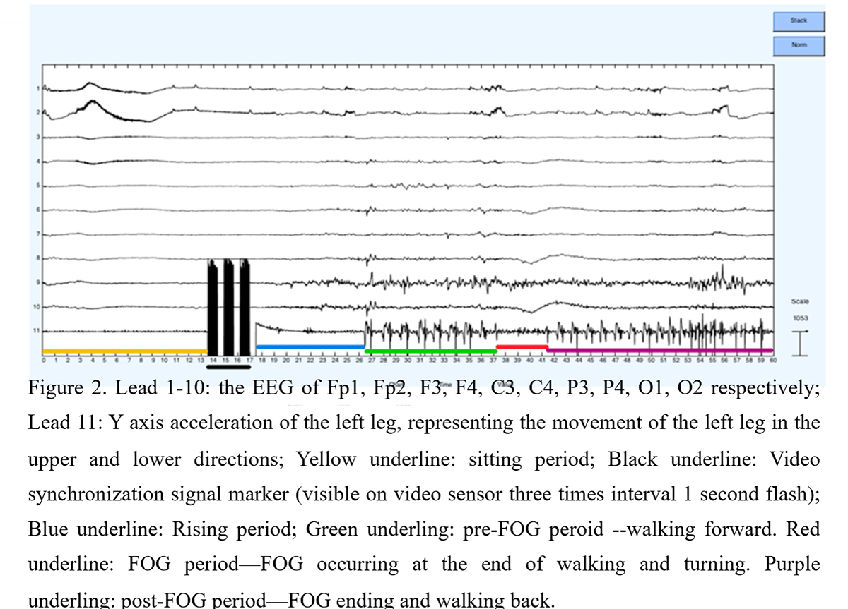
Method: A patient diagnosed with idiopathic PD with significant FOG, was assessed by video-recorded TUG task in the ”MED off” state following overnight withdrawal of dopaminergic therapy, using wearable Gai-EEG (figure 1) to simultaneously record the three-axis acceleration and scalp EEG. FOG onset during turning was identified and detected by the acceleration recording,the time-frequency analysis of EEG data was applied to investigate the dynamic EEG changes in real time, comparing episodes of pre, during and post FOG.
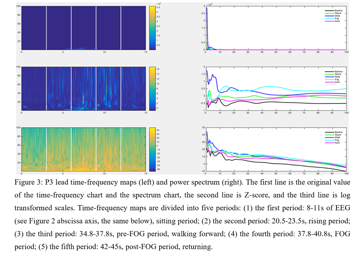
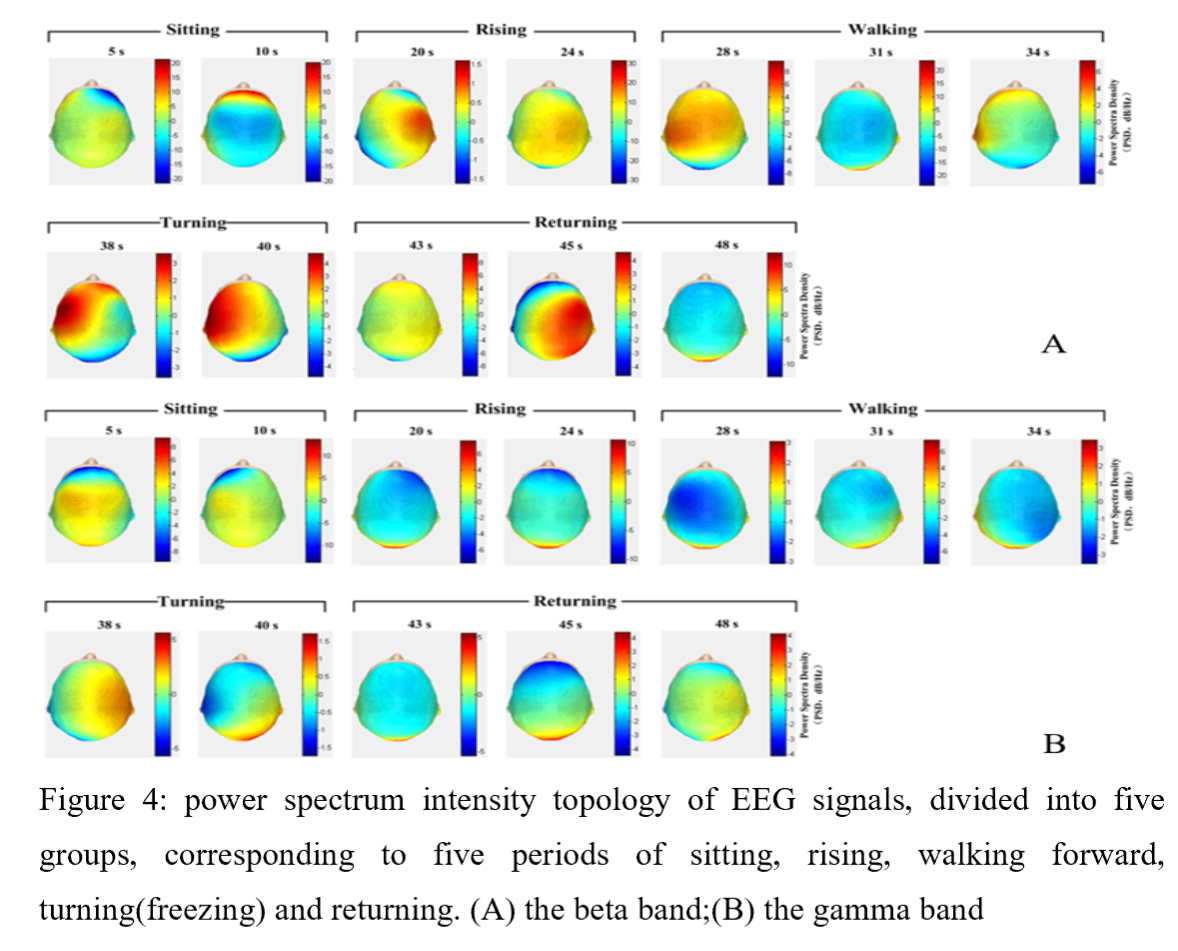
Results: We can confirm the FOG onset and ending time point during turning based on the acceleration recording, and divide the TUG process into five periods: sitting, rising, walking forward(pre-FOG), turning(FOG) and returning(post-FOG)(figure 2). High energy in low gamma and beta bands were found in the partial region during pre-FOG and FOG periods, the energy peak of the beta band were also found during the pre-FOG and FOG periods in the power spectrum, and the beta energy spectrum intensity in pre-FOG period was significant higher than that of in the post-FOG(figure 3). The increased power spectrum intensity of beta band was found in the left frontal-parietal region at the beginning of pre-FOG and the whole FOG period , and also found in the right frontal parietal region during the post-FOG peroid(figure 4A), which not found in gamma band (figure 4B).
Conclusion: We found the partial region was more affected during a turning FOG, significant changes in beta band were found before FOG occurring when the patient was about to execute turning movements. It’s able to reflect actual gait planning whilst turning, which may provide valuable information for the prediction of FOG.
To cite this abstract in AMA style:
J. Li, Y. Zhang. Time-Frequency analysis of Gait-EEG signals for freezing of gait in a PD patient [abstract]. Mov Disord. 2019; 34 (suppl 2). https://www.mdsabstracts.org/abstract/time-frequency-analysis-of-gait-eeg-signals-for-freezing-of-gait-in-a-pd-patient/. Accessed February 5, 2026.« Back to 2019 International Congress
MDS Abstracts - https://www.mdsabstracts.org/abstract/time-frequency-analysis-of-gait-eeg-signals-for-freezing-of-gait-in-a-pd-patient/


