Session Information
Date: Tuesday, September 24, 2019
Session Title: Parkinsonisms and Parkinson-Plus
Session Time: 1:45pm-3:15pm
Location: Agora 3 West, Level 3
Objective: Describe 4 cases of Parkinson’s disease (PD) patients with residual tremor post deep brain stimulation of subthalamic nucleus (DBS-STN) that underwent repositioning of electrodes posteriorly in the STN with subsequent clinical improvement of tremor.
Background: DBS-STN is a standard therapeutic option in PD with excellent short- and long-term clinical results. Optimal DBS site for PD patients with tremor, bradykinesia and rigidity appear to correspond to different areas in the motor STN. Stimulation of posterior STN is most effective for tremor as thought to be connected to the motor cortex M1. Whereas for rigidity and bradykinesia stimulation of mid STN connecting to SMA and PFMC, showed highest improvements [1].
Method: Retrospectively reviewed 4 cases with residual tremor after initial DBS-STN selected for repositioning of electrodes in the STN. Lead verification and repositioning was confirmed with MRI of brain. Pre-operative high resolution T1 and T2 weight MRI images were co-registered to initial and revision post-operative CT with LeadDBS Matlab Toolbox[2]. The co-registered scans were nonlinearly transformed into an MNI space atlas for visualization of patient data within a single reference brain atlas[3].
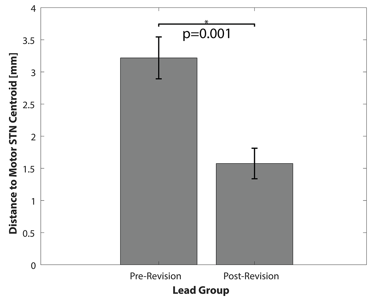
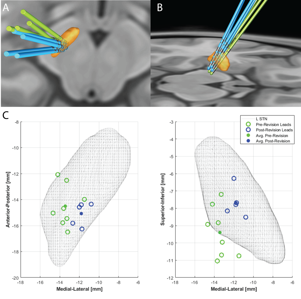
Results: In all 4 cases, patients had significant improvement post initial DBS , however had suboptimal tremor control and stimulation side effects. UPDRS OFF meds was 43+/- 3, UPDRS ON 25+/- 3. Post DBS UPDRS score improved 58% (UPDRS DBS ON=16.5). All cases had refractory rest tremor that was not controlled in spite of multiple programing attempts. Post repositioning, all patients had improvement in tremor control. The distances of the pre-revision and post-revision leads to the centroid of the motor STN were significantly closer with mean decease in distance of 1.64.mm [Figure 1]. In all cases, electrodes were repositioned on average superior, medial, and posterior in STN compared to initial electrodes [Figure 2].
Conclusion: Our case series supports that stimulation of the posterior/superior STN area is effective for tremor control.
References: 1. Akram et al. Subthalamic deep brain stimulation sweet spots and hyperdirect cortical connectivity in Parkinson’s disease, Neuroimage. 2017 Sep;158:332-345 2. Andreas Horn, Andrea Kühn. Lead-DBS: A toolbox for deep brain stimulation electrode localizations and visualizations. NeuroImage, 2014. 3. Ewert, S., Plettig, P., Li, N., Chakravarty, M. M., Collins, D. L., Herrington, T. M., et al. Toward defining deep brain stimulation targets in MNI space: A subcortical atlas based on multimodal MRI, histology and structural connectivity. NeuroImage 2017.
To cite this abstract in AMA style:
T. Stiep, A. Diaz, I. Cajigas, J. Jagid, C. Luca. The STN Sweet Spot for Tremor Control in Parkinson’s Disease [abstract]. Mov Disord. 2019; 34 (suppl 2). https://www.mdsabstracts.org/abstract/the-stn-sweet-spot-for-tremor-control-in-parkinsons-disease/. Accessed December 14, 2025.« Back to 2019 International Congress
MDS Abstracts - https://www.mdsabstracts.org/abstract/the-stn-sweet-spot-for-tremor-control-in-parkinsons-disease/


