Category: Parkinson's Disease: Non-Motor Symptoms
Objective: Our goal was to investigate retinal structural changes as well as changes in visual cortex in PD patients with suppression deficiency of VOR and gait disorders.
Background: The development of structural changes in retina and cortical visual centers due to dopamine deficiency, as well as disintegration between the visual and vestibular systems, is the basis for gait disorders in PD. Therefore, the study of vestibuloocular reflex (VOR) suppression provides the valuable information regarding the interaction of the above-mentioned systems and can be used in risk assessment of falls in PD patients
Method: 50 PD patients aged 45-65 years, as well as 30 healthy people were examined. We used optical coherence tomography, MRI voxel-based morphometry and Functional Gait Assessment scale. Horizontal and vertical VOR was studied by videonystagmography with sinusoidal rotary chair stimulation in dark (test 1) and fixed gaze on head-fixed target (test 2) in horizontal and vertical plane.
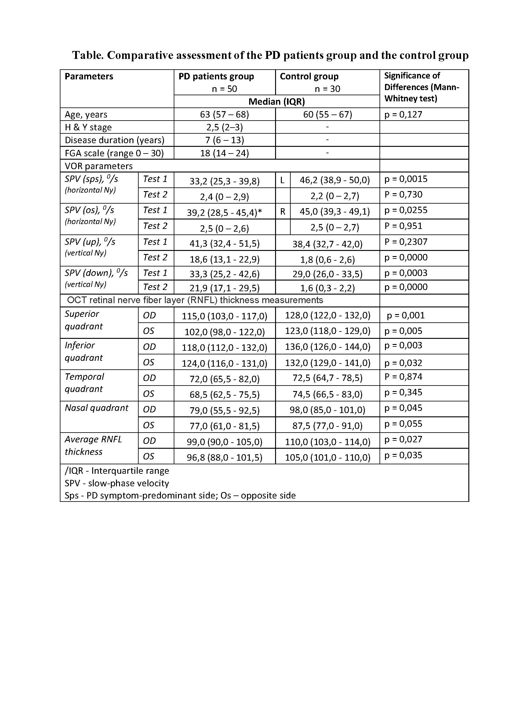
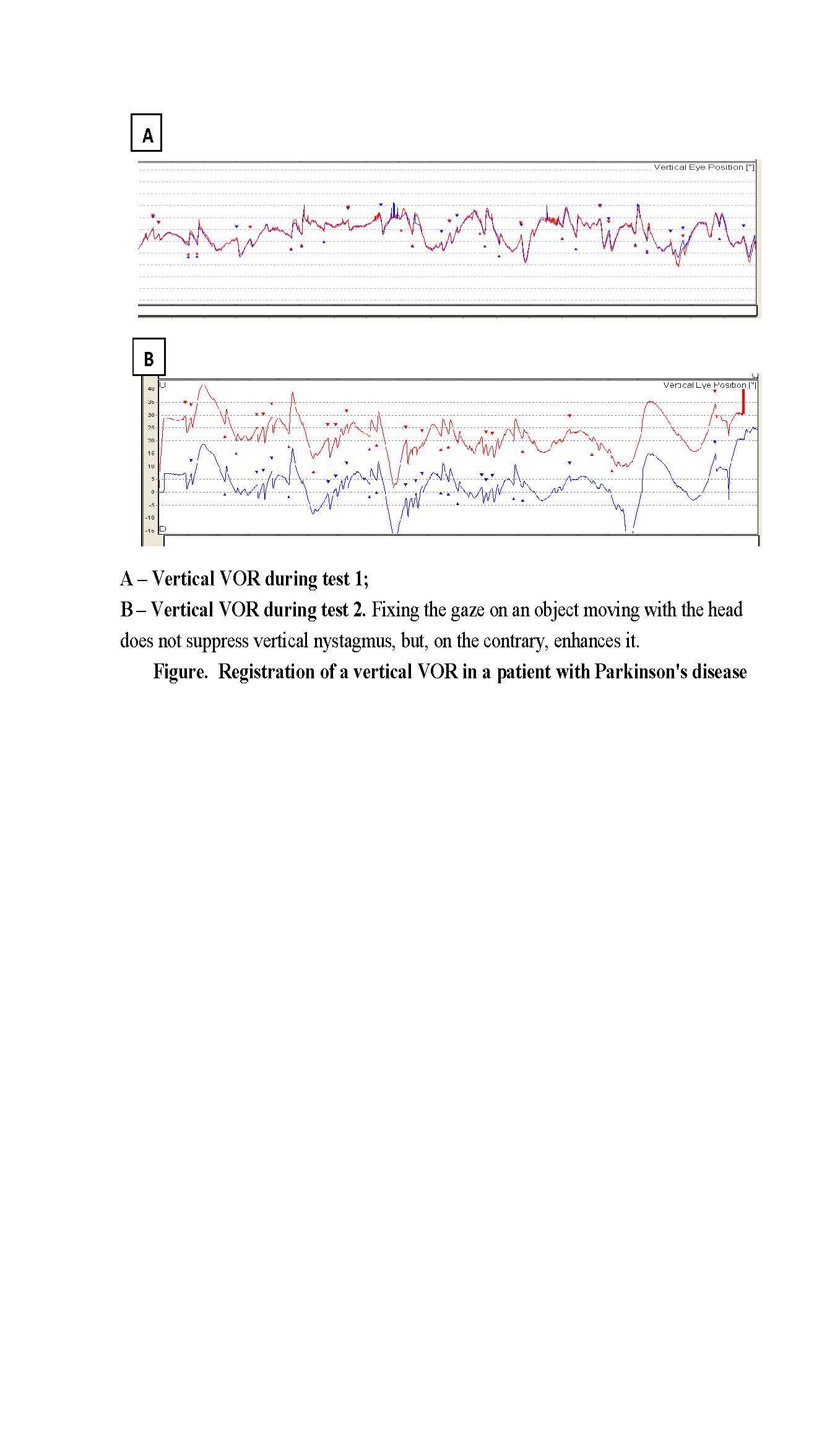
Results: The results showed that the average RNFL thickness in PD was reduced significantly, especially in the superior and inferior quadrant compared with healthy controls (Table 1). Same as the control group, the PDpatients showed complete suppression of horizontal nystagmus during rotation in horizontal plane, but they were unable to suppress vertical nystagmus during rotation in vertical plane (Figure). MRI revealed a decrease in the brain structures volume involved in visual information processing in PD.
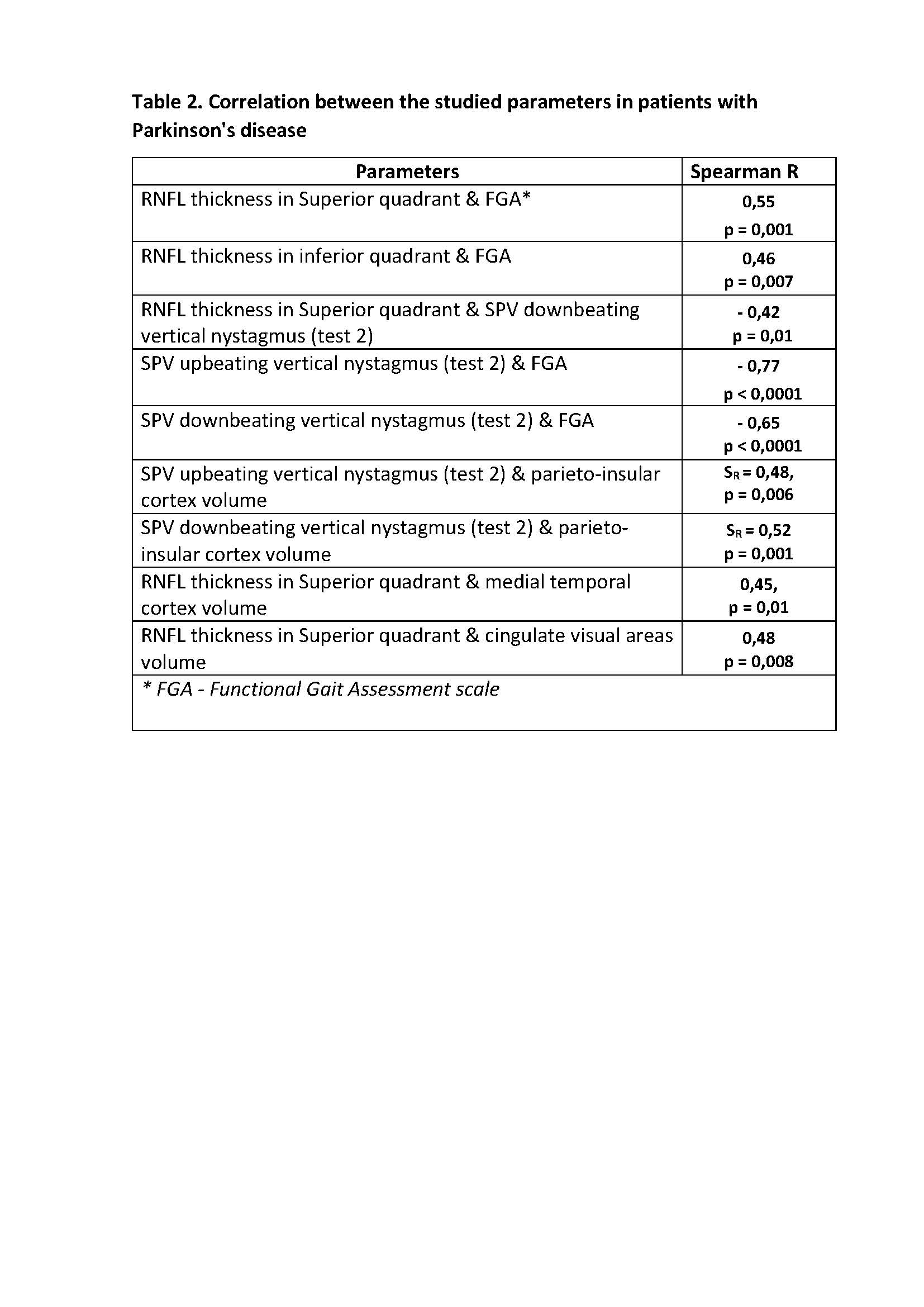
RNFL thickness in the superior and inferior quadrants correlated with gait disorders, as well as the suppression deficiency of vertical VOR. Additionally, the suppression deficiency of vertical VOR correlated with the decrease in the parieto-insular cortex volume, and the thickness in superior quadrant correlated with the decrease in the medial temporal area and cingulate visual areas volume (table 2).
Conclusion: The link between structural disorders in superior and inferior regions of the retina and gait abnormalities in PD reflects the critical role of visual information for gait. Besides the retinal change and visual cortical area reduction, the PDpatients developed inability to process properly vestibular information in interaction with visual stimuli. So, analysing the association of VOR suppression with retinal structural abnormality can assess the risks of falls in PD.
To cite this abstract in AMA style:
O. Alenikova, O. Davydova, N. Alenikov. The role of structural changes in the retina and the vertical vestibulo-ocular reflex abnormality in gait disorders in Parkinson`s disease [abstract]. Mov Disord. 2023; 38 (suppl 1). https://www.mdsabstracts.org/abstract/the-role-of-structural-changes-in-the-retina-and-the-vertical-vestibulo-ocular-reflex-abnormality-in-gait-disorders-in-parkinsons-disease/. Accessed September 12, 2025.« Back to 2023 International Congress
MDS Abstracts - https://www.mdsabstracts.org/abstract/the-role-of-structural-changes-in-the-retina-and-the-vertical-vestibulo-ocular-reflex-abnormality-in-gait-disorders-in-parkinsons-disease/


