Session Information
Date: Thursday, June 8, 2017
Session Title: Parkinson's Disease: Neuroimaging And Neurophysiology
Session Time: 1:15pm-2:45pm
Location: Exhibit Hall C
Objective: The role of MRI and DAT scan in Vascular Parkinsonism: A Case Report
Background: DaTscan has become a widely used clinical tool to help clinicians with challenging diagnoses. DaTscan can help distinguish idiopathic Parkinson Disease (PD) from secondary forms of Parkinsonism such as vascular Parkinsonism (VD). The integrity of the nigrostriatal dopamine pathway can be assessed using single photon emission computed tomography (SPECT) with dopamine transporter (DaT) radioligands. DaTscan would be abnormal in PD and normal in VD. While a meta-analysis by Brigo showed high sensitivity and specificity (above 80%) in differentiating PD and VD using DaTscan, reliance on DaTscan alone may be misleading in certain cases.
Methods: Case study
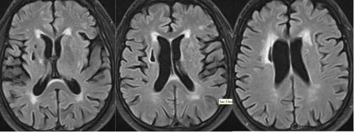
Results: 65 year-old man with atrial fibrillation on warfarin, coronary artery disease and carotid artery stenosis with right carotid endarterectomy presented with falls and left hand tremors for 6 months. Exam showed moderate bradykinesia on the left side and mild left hand action tremor with decreased bilateral arm swings, decreased stride length, difficulty with turns and postural instability. There were no pyramidal signs. DaTscan (figure 1) showed reduced dopamine transporter uptake bilaterally, right greater than left. His tremor improved with levodopa but his gait did not. Subsequent MRI brain (figure 2) showed extensive microvascular white matter disease and a large right basal ganglia infarct extending into the right corona radiata. Our patient is presumed to have VP and not PD. VP tends to occur at an older age with sudden onset, prominent lower body involvement, less response to levodopa, gait and postural instability, urinary incontinence, pyramidal signs and dementia compared to PD.
Conclusions: Although DaTscan has been shown to have high sensitivity and specificity by many studies, the test has limitations because in most studies the gold standard used is the clinical diagnosis made by a movement disorders neurologist and not a neuropathological confirmation. Therefore, the true diagnosis is unknown; false positive or false negative scans can occur, which highlights the needs for integration of various clinical findings and imaging tools (MRI and DaTscan). This case illustrates an abnormal DaTscan in a patient with VD.
References: Brigo F, et al. [123I]FP-CIT SPECT (DaTscan) may be a useful tool to differentiate between Parkinson’s disease and vascular or drug-induced parkinsonisms: A meta-analysis. Eur J Neurol 2014;21:1369-1376.
Vale TC, et al. Clinicoradiological comparison between vascular parkinsonism and Parkinson’s disease. J Neurol Neurosurg Psychiatry. 2015 May;86(5):547-53. Epub 2014 Jul 8.
To cite this abstract in AMA style:
A. Tran, M. Amin, R. Burns. The Role of MRI and DaTscan in Vascular Parkinsonism: A Case Report [abstract]. Mov Disord. 2017; 32 (suppl 2). https://www.mdsabstracts.org/abstract/the-role-of-mri-and-datscan-in-vascular-parkinsonism-a-case-report/. Accessed October 22, 2025.« Back to 2017 International Congress
MDS Abstracts - https://www.mdsabstracts.org/abstract/the-role-of-mri-and-datscan-in-vascular-parkinsonism-a-case-report/


