Category: Parkinson's Disease: Neuroimaging
Objective: Study the effect of propranolol on (stress-related increase in) tremor power and on tremor-related activity in the cerebello-thalamo-cortical (CTC) circuit in Parkinson’s (PD) patients.
Background: Previous work showed that PD resting tremor is initiated in the basal ganglia and amplified in the CTC circuit[1]. Since stress worsens tremor, the noradrenergic (NA) stress system might amplify tremor. We confirmed that stress increased tremor through bottom-up thalamus excitation[2], but it remains unclear how this is mediated by NA activity.
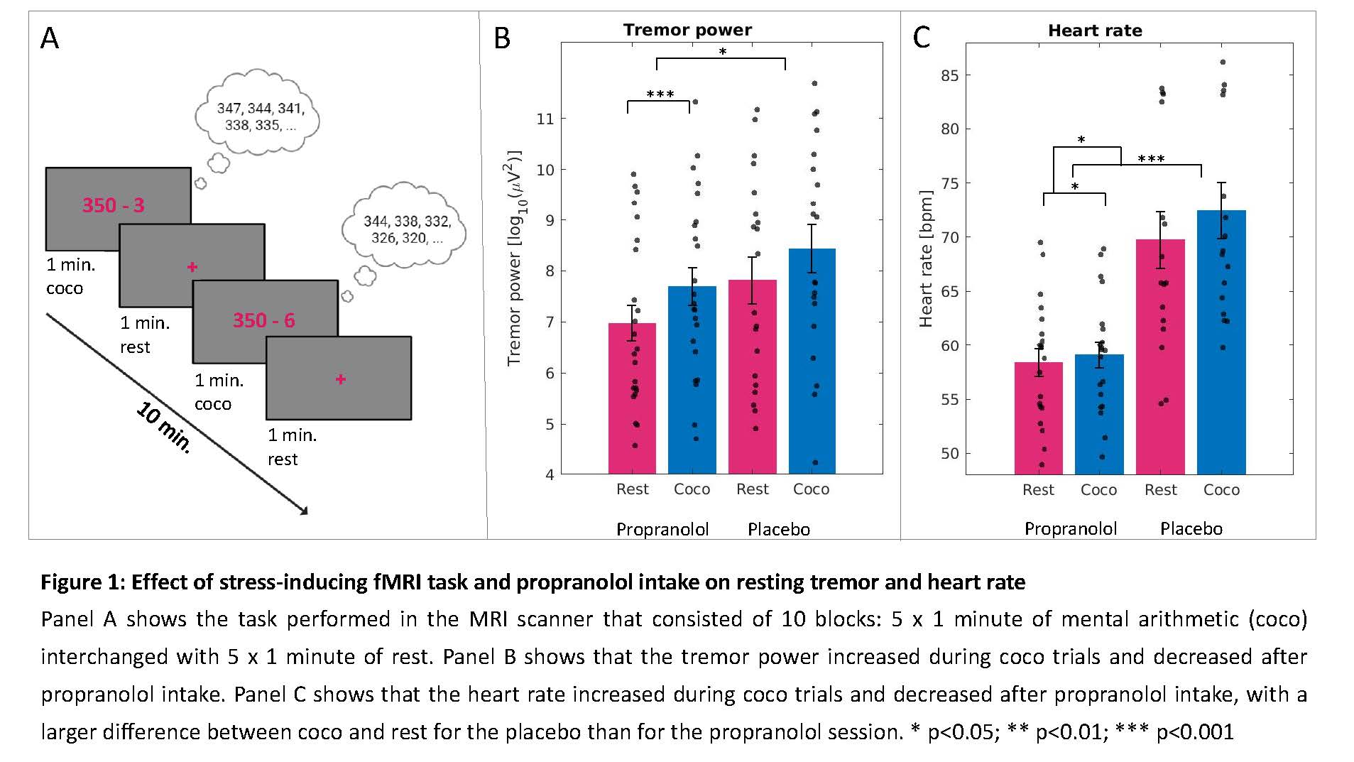
Method: In a cross-over double-blind intervention study, 25 PD patients with tremor underwent fMRI twice, once receiving propranolol (40mg, oral) and once placebo (counterbalanced). The NA system was activated with a stress-inducing cognitive-coactivation (coco) task during both sessions consisting of alternating mental arithmetic and rest blocks [figure1]. We measured brain activity (fMRI), tremor power (EMG+accelerometry), pupil diameter (eye tracking) and heart rate (HR). We tested for effects of DRUG (propranolol vs. placebo) and TRIAL (coco vs. rest) on both task-related and tremor-related brain activity, correcting for multiple comparisons (p<.05 FWE) at the whole brain for task-related effects and in the CTC circuit for tremor-related effects.
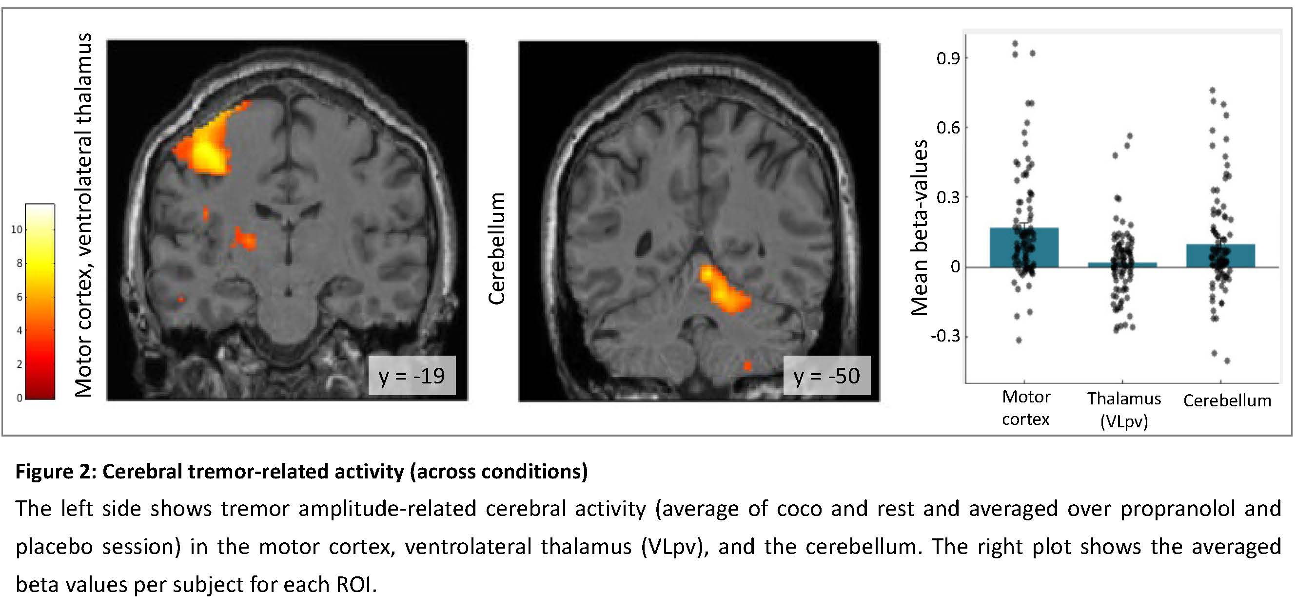
Results: Tremor power was increased by coco (p=.001) and decreased by propranolol (p=.02) (Fig. 1B). HR increased during coco trials (p=.03), and was lowered by propranolol (p<.001), with a larger coco-rest difference for placebo than for propranolol (p=.04) (Fig. 1C). Whole brain analysis showed coco-related activity in the SMA [max t(89)=11.3; p<.001], precentral sulcus [max t(89)=9.3; p<.001], sup. and inf. parietal lobe [max t(89)=9.9; p<.001], insula [max t(89)=7.2; p<.001], and cerebellum [max t(89)=10.0; p<.001]. Tremor-amplitude related activity was found in the ipsilateral cerebellum [t(89)=4.9; p<.001] and contralaterally in motor cortex [t(89)=7.7; p<.001], and ventrolateral thalamus [t(89)=3.4; p=.02] (Fig. 2). There were no main effects of DRUG and no DRUG*TRIAL interactions.
Conclusion: Activation of the NA system with a cognitive task increased tremor power, while suppression of the NA system with propranolol lowered tremor power. Cognitive load increased activity in a cognitive control network, tremor-related activity was observed in a CTC network. We will test how propranolol influences the interaction between these networks using dynamic causal modelling.
References: [1] Helmich, R. C., Hallett, M., Deuschl, G., Toni, I. & Bloem, B. R. Cerebral causes and consequences of parkinsonian resting tremor: a tale of two circuits? Brain 135, 3206-3226, doi:10.1093/brain/aws023 (2012).
[2] Dirkx, M. F. et al. Cognitive load amplifies Parkinson’s tremor through excitatory network influences onto the thalamus. Brain 143, 1498–1511, doi:10.1093/brain/awaa083 (2020).
To cite this abstract in AMA style:
A. Heide, M. Wessel, D. Papadopetraki, D. Geurts, T. van Prooije, F. Gommans, R. Helmich. The noradrenergic basis of Parkinson’s tremor [abstract]. Mov Disord. 2022; 37 (suppl 2). https://www.mdsabstracts.org/abstract/the-noradrenergic-basis-of-parkinsons-tremor/. Accessed April 18, 2025.« Back to 2022 International Congress
MDS Abstracts - https://www.mdsabstracts.org/abstract/the-noradrenergic-basis-of-parkinsons-tremor/


