Category: Parkinson's Disease: Neuroimaging
Objective: To improve cerebral vascular visibility using an ultra-small super-paramagnetic iron oxide (USPIO) contrast agent and identify vascular abnormalities in patients with Parkinson’s disease (PD).
Background: Developmental venous anomalies (DVAs) are the most common vascular malformations with a prevalence of up to 5%.1,2The most common location is the frontal lobe and least likely is the basal ganglia (BG).3 Although DVAs drain normal brain parenchyma, changes such as regional brain atrophy and white matter lesions due to impaired perfusion as a result of chronic venous hypertension have been reported.2,3 Cerebral ischemia triggers a cascade of events that includes oxidative stress and inflammation that promotes neurodegeneration.4,5 DVAs are usually asymptomatic, but atypical DVAs have been reported to cause neurological symptoms. Their association with PD, however, has never been explored.
Method: Thirty patients with PD were enrolled. All participants were scanned at 3T utilizing MICRO (Microvascular In-vivo Contrast Revealed Origins) imaging, which included 3D high-resolution (0.22×0.44×1 mm3) susceptibility weighted imaging (SWI) data acquired before, during and after the infusion of Ferumoxytol at a dose of 4mg/kg6. The imaging parameters were: TEs = 9.5, 16, 22.5 ms, TR = 31 ms, flip angle = 12° and bandwidth = 177 Hz/pxl. SWI images were generated by homodyne high pass filtering (filter size = 96 × 96) the phase images. A phase mask was then created from the high pass filtered phase data and multiplied into the original magnitude images four times.
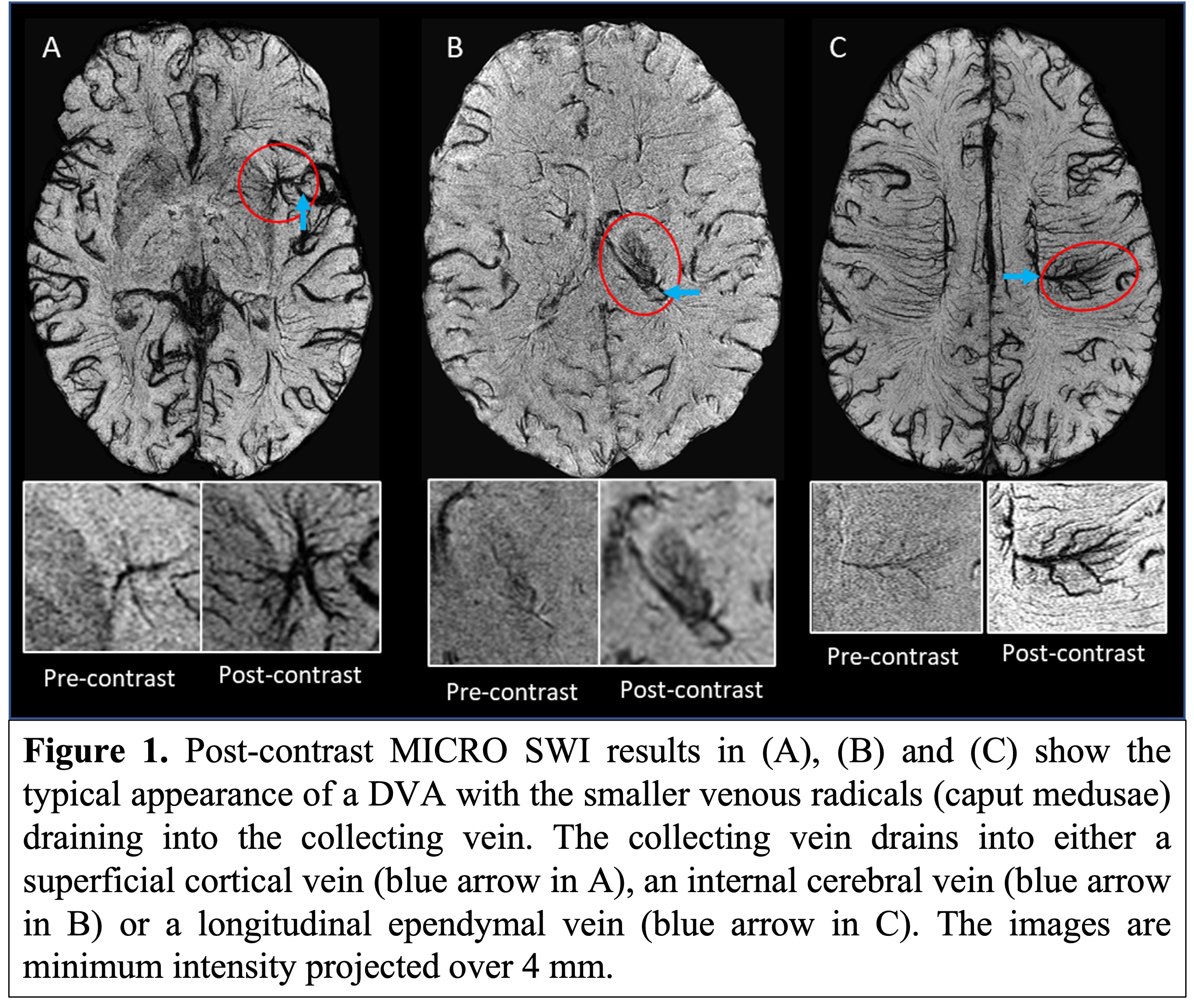
Results: DVAs were found in 5 (16.6%) patients. All DVAs were supratentorial (ST) and located in the thalamus (n=1), putamen (n=2), lentiform nucleus (n=1) and the fronto-parietal region (n=1). The drainage of the DVAs was into the deep ST veins (Figure 1B) except in one case where it drained into a superficial ST vein (Figure 1A). None were associated with any other pathologies.
Conclusion: MICRO imaging enhances the underlying vascularity usually undetectable on non-contrast SWI and has the potential to study the vascular etiology of PD. DVAs were found to be more common in patients with PD than the general population. The high vulnerability of the BG to chronic ischemia facilitated by the presence of poor flow in a DVA could be contributory to PD pathogenesis.
References: 1. Rinaldo L, Lanzino G, Flemming KD, Krings T, Brinjikji W. Symptomatic developmental venous anomalies. Acta Neurochirurgica. 2020 May;162(5):1115-25.
2. Lasjaunias P, Burrows P, Planet C. Developmental venous anomalies (DVA): the so-called venous angioma. Neurosurgical review. 1986 Sep;9(3):233-42.
3. San Millán Ruíz D, Delavelle J, Yilmaz H, Gailloud P, Piovan E, Bertramello A, Pizzini F, Rüfenacht DA. Parenchymal abnormalities associated with developmental venous anomalies. Neuroradiology. 2007 Dec;49(12):987-95.
4. Kim T, Vemuganti R. Mechanisms of Parkinson’s disease-related proteins in mediating secondary brain damage after cerebral ischemia. Journal of Cerebral Blood Flow & Metabolism. 2017 Jun;37(6):1910-26.
5. Lin B, Levy S, Raval AP, Perez-Pinzon MA, Defazio RA. Forebrain ischemia triggers GABAergic system degeneration in substantia nigra at chronic stages in rats. Cardiovascular Psychiatry and Neurology. 2010;2010.
6. Buch S, Wang Y, Park MG, Jella PK, Hu J, Chen Y, Shah K, Ge Y, Haacke EM. Subvoxel vascular imaging of the midbrain using USPIO-Enhanced MRI. Neuroimage. 2020 Oct 15;220:117106.
To cite this abstract in AMA style:
S. Sharma, S. Buch, D. Reese, T. Wade, G. Prado Miranda, M. Haacke, C. Tzoulis, M. Jog. The High Prevalence of Developmental Vascular Anomalies in Parkinson’s Disease using USPIO-based MICRO MRI [abstract]. Mov Disord. 2023; 38 (suppl 1). https://www.mdsabstracts.org/abstract/the-high-prevalence-of-developmental-vascular-anomalies-in-parkinsons-disease-using-uspio-based-micro-mri/. Accessed January 17, 2026.« Back to 2023 International Congress
MDS Abstracts - https://www.mdsabstracts.org/abstract/the-high-prevalence-of-developmental-vascular-anomalies-in-parkinsons-disease-using-uspio-based-micro-mri/

