Category: Tremor
Objective: To assess the feasibility of MRgFUS lesioning in the context of prior hemicraniotomy
Background: Targeted stereotactic lesioning, using MRgFUS, is an effective treatment option for many tremor disorders. Whilst the degree of treatment success may vary, depending on both disease and patient related factors, both ventralis intermediate nucleus (VIM) and posterior subthalamic area (PSA) lesioning are safe and efficacious targets for achieving satisfactory tremor control. As the indications for MRgFUS lesioning continue to be explored, the presence of prior craniotomy is a possible barrier to accurate lesioning.
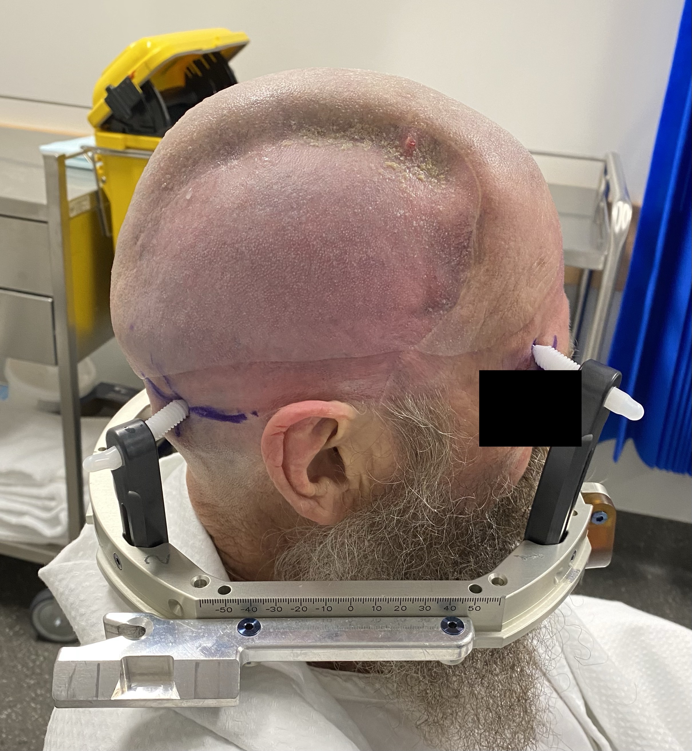
Method: A 68-year-old left-hand-dominant male, with a history of right-sided hemicraniotomy, underwent MRgFUS right VIM thalamotomy and PSA lesioning for the treatment of a severe upper limb posttraumatic tremor disorder. The hemicraniotomy had been performed in the context of an ICH sustained following a motor vehicle accident, forty years prior (figure 1). Baseline MR imaging noted gliotic change in the anterior right frontal and temporal lobes consistent with previous traumatic brain injury. The calculated SDR was 0.51. Areas of defect where the skull edges were not in direct opposition were excluded as ‘no-pass’ regions but the craniotomy flap was not excluded. A total of 887 elements were engaged with remaining skull surface area of 323cm2. A total of five therapeutic sonications of at least 54°C were administered.
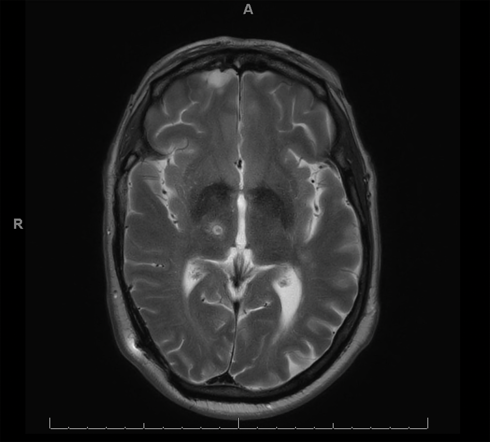
Results: At baseline, the total Clinical Rating Scale in Essential Tremor (CRST) score was 90 with a subset Hand Tremor Score (HTS) of 27. Despite immediate improvement, the tremor recurred at 1 month with a total CRST of 79 and HTS of 25. The patient reported mild non-bothersome taste disturbance and no other adverse events. Post procedure MR imaging revealed two well placed lesions in the right VIM (figure 2) and PSA.
Conclusion: This case, in addition to a previously published report of MRgFUS following craniotomy in essential tremor1, affirms the feasibility of this treatment modality in individuals with a skull defect secondary to prior neurosurgery. In this case, the tremor recurred despite initial improvement, and this is possibly a reflection of the complex network abnormalities seen in posttraumatic tremor disorders. However, in the presence a craniotomy flap and skull defect, MRgFUS achieved accurately placed lesions in the VIM and PSA.
References: 1. Wathen, Connor MD*; Yang, Andrew I. MD, MS*; Hitti, Frederick L. MD, PhD*; Henry, Lenora MS‡; Chaibainou, Hanane CRNP, MSN*; Baltuch, Gordon H. MD, PhD*. Feasibility of Magnetic Resonance–Guided Focused Ultrasound Thalamotomy for Essential Tremor in the Setting of Prior Craniotomy. Operative Neurosurgery 22(2):p 61-65, February 2022. | DOI: 10.1227/ONS.0000000000000012
To cite this abstract in AMA style:
J. Maamary, J. Peters, Y. Barnett, B. Jonker, S. Tisch. The feasibility of MRI guided focused ultrasound for posttraumatic tremor with prior hemicraniotomy and skull defect [abstract]. Mov Disord. 2023; 38 (suppl 1). https://www.mdsabstracts.org/abstract/the-feasibility-of-mri-guided-focused-ultrasound-for-posttraumatic-tremor-with-prior-hemicraniotomy-and-skull-defect/. Accessed May 11, 2025.« Back to 2023 International Congress
MDS Abstracts - https://www.mdsabstracts.org/abstract/the-feasibility-of-mri-guided-focused-ultrasound-for-posttraumatic-tremor-with-prior-hemicraniotomy-and-skull-defect/


