Category: Parkinson's Disease: Neuroimaging
Objective: We aim to decode the neuroimaging features of LC in Parkinson’s tremor.
Background: Tremor is the primary symptom of Parkinson’s disease (PD) and exhibits complex pathophysiology potentially implicating the locus coeruleus (LC) noradrenergic system. However, the role and mechanisms of LC in Parkinson’s tremor still remain unclear.
Method: Forty-eight PD patients with tremor dominant (TD), 72 PD patients with postural instability/gait disorder (PIGD), and 83 age-matched healthy controls (HCs) scanned magnetic resonance imaging (MRI). Neuromelanin sensitive MRI (NM-MRI) and resting-state functional MRI (rs-fMRI) were used to investigate the degeneration of LC neurons and the effective connectivity (EC) between LC and cerebello-thalamo-cortical (CTC) circuit based on spectral dynamic causal modeling (spDCM) in rs-fMRI on the more-affected and less-affected side. We focused on the 3 nodes of the CTC circuit: ventral intermediate nucleus (VIM), motor cortex (MC), and cerebellum (CBLM). Furthermore, we analyze the associations between the imaging alterations of LC and the clinical evaluations to elucidate the pathological underpinnings of Parkinson’s tremor.
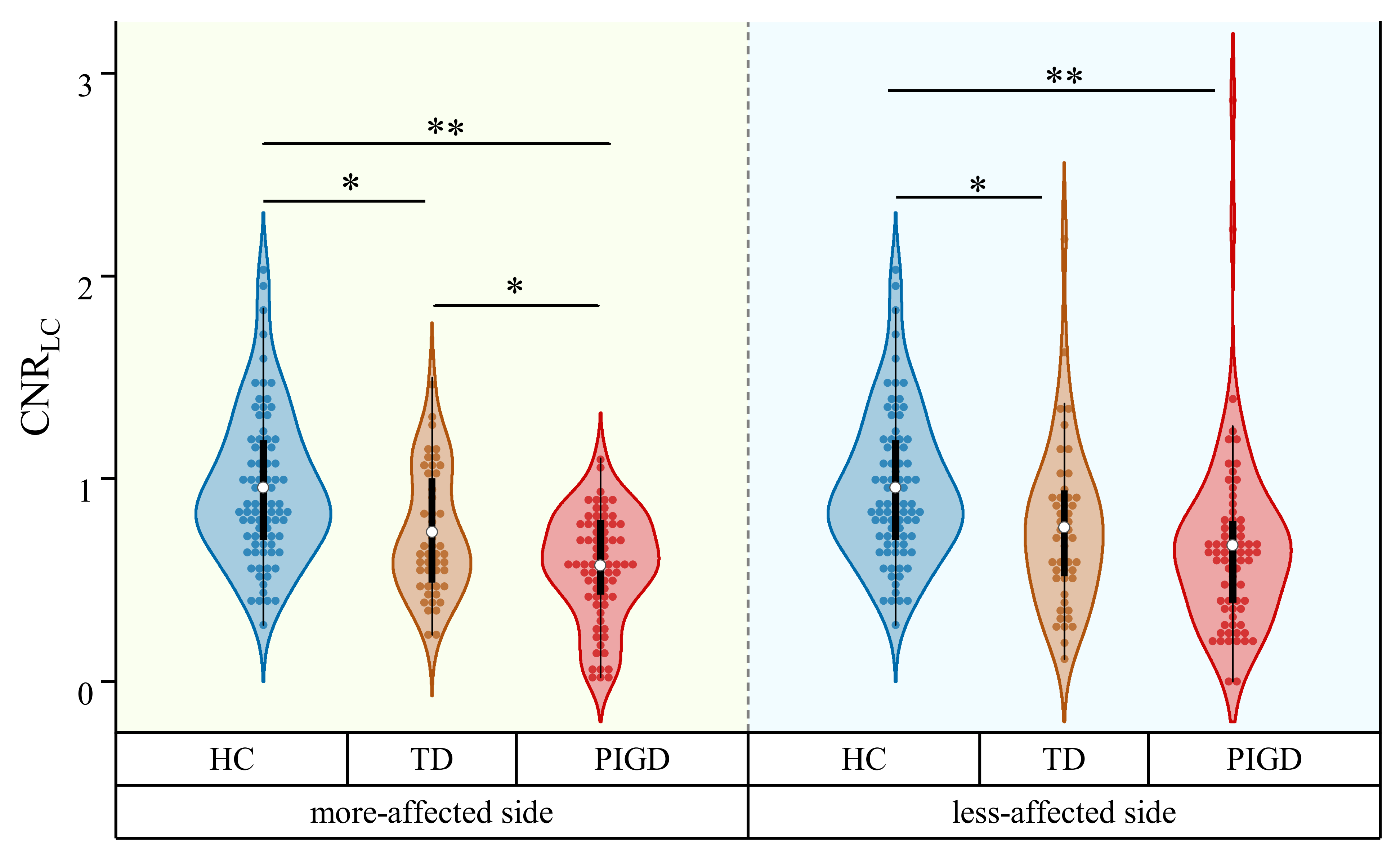
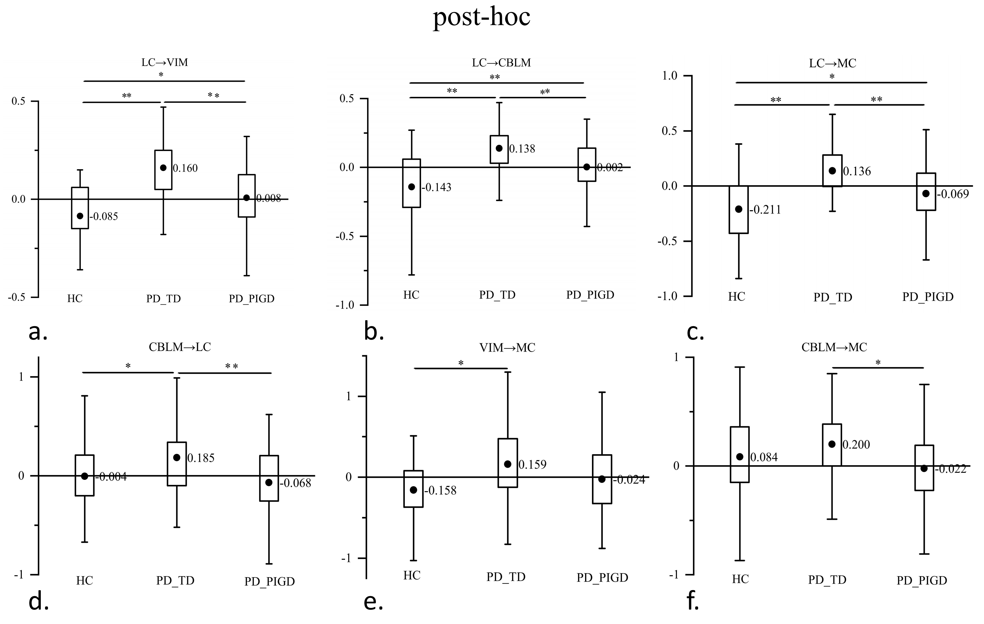
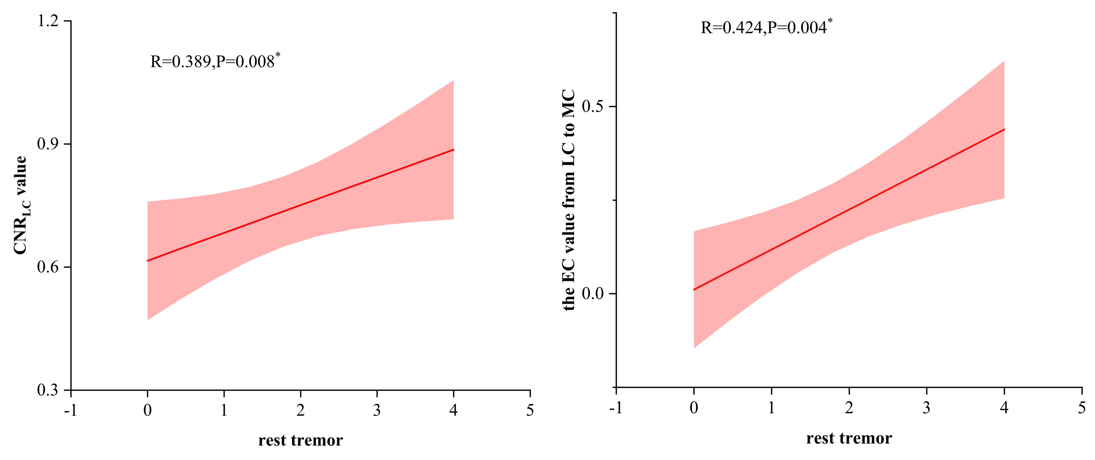
Results: We found that the contrast-to-noise ratio of LC (CNRLC) value was decreased bilaterally in the patients with TD and PIGD compared to HCs. The patients with TD showed significantly higher CNRLC value compared to the patients with PIGD on the more-affected side (Figure 1). Compared to HCs and the patients with PIGD, the EC value from LC to the 3 nodes of CTC circuit on the more-affected side increased significantly in the patients with TD . Within the CTC circuit, the EC value from VIM to MC increased significantly in the patients with TD than HCs, and the EC value from CBLM to MC preserved relatively than the patients with PIGD (Figure 2) (p<0.0125, LSD corrected). In addition, we found that the CNRLC value and the EC value from LC to MC on the more-affected side positively correlated with the amplitude of the rest tremor in the patients with TD (R=0.389, P=0.008; R=0.424, P=0.004, respectively) (Figure 3).
Conclusion: Our findings suggested that LC structural and functional impairment are uncoupled. which may be a key pathological mechanism for the Parkinson’s tremor. This study provides a neuroimaging rationale for the development of therapeutic strategies targeting LC.
Fig. 1 The CNRLC value of LC in participants.
Fig. 2 The EC value between LC and CTC circuit
Fig. 3 LC imaging signs correlated with tremor
To cite this abstract in AMA style:
JY. Sun. the Degeneration of Locus Coeruleus and its Enhanced Causal Relationship to Neural Circuit in Parkinson’s Tremor [abstract]. Mov Disord. 2024; 39 (suppl 1). https://www.mdsabstracts.org/abstract/the-degeneration-of-locus-coeruleus-and-its-enhanced-causal-relationship-to-neural-circuit-in-parkinsons-tremor/. Accessed April 20, 2025.« Back to 2024 International Congress
MDS Abstracts - https://www.mdsabstracts.org/abstract/the-degeneration-of-locus-coeruleus-and-its-enhanced-causal-relationship-to-neural-circuit-in-parkinsons-tremor/


