Category: Parkinson's Disease: Neuroimaging
Objective: Determine whether MRI can identify differences in hippocampal volume between patients with Lewy body disorders with and without Alzheimer’s copathology, and whether these differences explain variation in verbal memory performance.
Background: Lewy body disorders (LBD) include Parkinson disease (PD), Parkinson disease dementia (PDD), and dementia with Lewy bodies (DLB). Around half of LBD patients have coexisting AD pathology (LBD+AD), which correlates with shorter time to dementia and death. Verbal memory (VM) impairment correlates with worse hippocampal tau pathology at autopsy in LBD. It remains unknown whether hippocampal tau burden in LBD+AD causes neurodegeneration detectable by in vivo structural MRI, and whether hippocampal atrophy explains VM impairment.
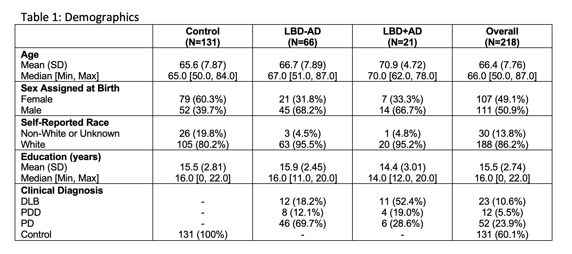
Method: We studied 87 patients with clinical diagnoses of LBD (PD, n = 52; PDD, n = 12; DLB, n = 23) with available T1 MRI and AD biomarkers or autopsy, and cognitively-normal controls (n = 131). AD copathology was defined hierarchically by: a) AD “intermediate” or “high” by ABC neuropathologic criteria; 2) positive amyloid PET; and 3) CSF total tau:β-amyloid1-42 ratio > 0.3 (autopsy-validated cutpoint). The Automated Segmentation of Hippocampal Subfields pipeline was applied to subjects’ T1 MRIs. Hippocampal volume (HV) was compared between controls, LBD-AD, and LBD+AD, and correlated with VM scores. Linear regression was used to test the association of AD copathology and HV (dependent variable), covarying for age, sex, education, and clinical diagnosis.
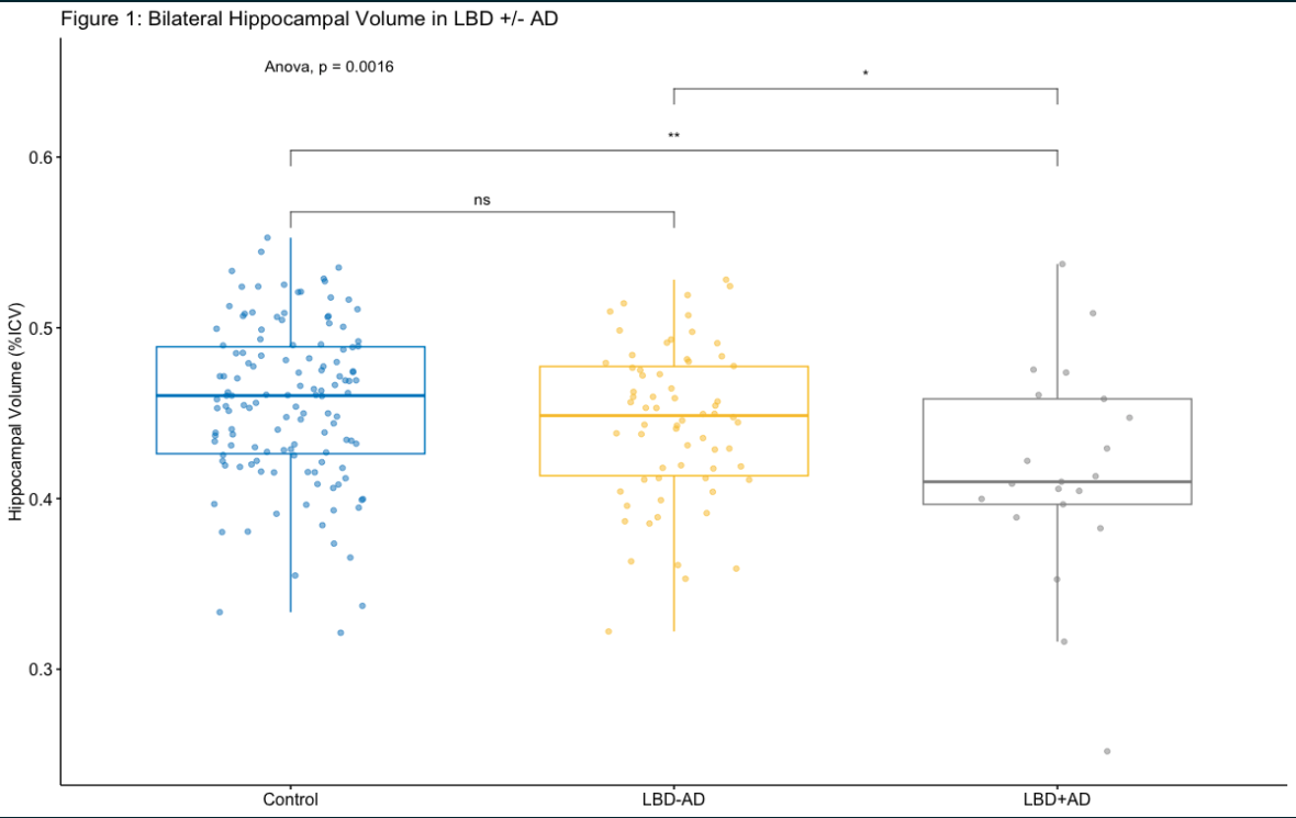
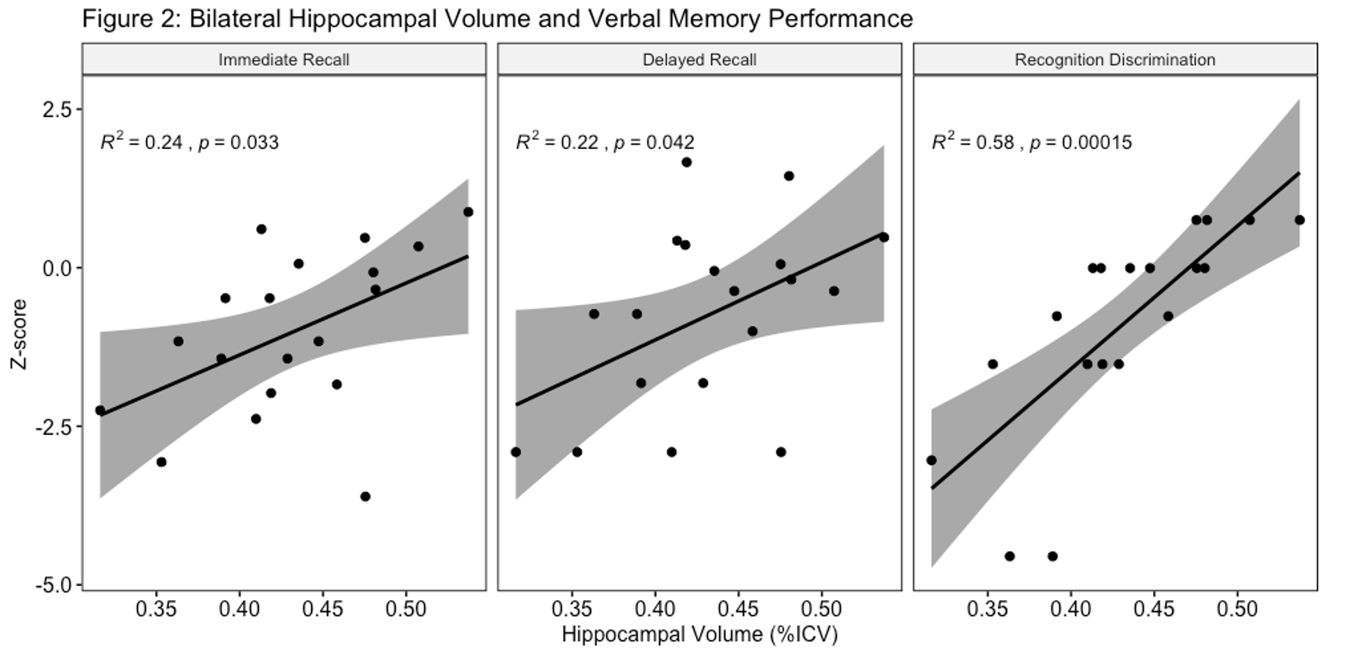
Results: Significant differences (ANOVA p=0.002) in bilateral HV (expressed as percentage of intracranial volume) were seen between groups (mean 0.456±0.046 for controls, 0.444±0.045 for LBD-AD, and 0.416±0.063 for LBD+AD). Tukey’s test showed significant differences between LBD+AD vs controls (p=0.001) and LBD+AD vs. LBD-AD (p=0.047). Linear regression showed a significant association of LBD+AD with HV (p=0.034). Among all LBD patients, HV positively correlated with VM (immediate recall: r2 = 0.24, p=0.033, delayed recall: r2 = 0.22, p=0.042, recognition discrimination r2 = 0.58, p<0.001).
Conclusion: In patients with LBD, AD copathology associates with greater hippocampal atrophy on T1 MRI, which correlates with greater verbal memory deficits. This supports the hypothesis that AD copathology impairs cognition in LBD via hippocampal neurodegeneration.
References: 1. Irwin, D.J., et al., Neuropathological and genetic correlates of survival and dementia onset in synucleinopathies: a retrospective analysis. Lancet Neurol, 2017. 16(1): p. 55-65.
2. Coughlin, D.G., et al., Hippocampal subfield pathologic burden in Lewy body diseases vs. Alzheimer’s disease. Neuropathology and Applied Neurobiology, 2020. 46(7): p. 707-721.
3. Irwin, D.J., et al., CSF tau and β-amyloid predict cerebral synucleinopathy in autopsied Lewy body disorders. Neurology, 2018. 90(12): p. e1038-e1046.
4. Xie, L., et al., Automated segmentation of medial temporal lobe subregions on in vivo T1-weighted MRI in early stages of Alzheimer’s disease. Hum Brain Mapp, 2019. 40(12): p. 3431-3451.
To cite this abstract in AMA style:
J. Cohen, J. Phillips, S. Das, E. Rhodes, K. Cousins, P. Yushkevich, D. Wolk, D. Weintraub, D. Irwin, C. Mcmillan. T1 MRI reveals differential hippocampal atrophy in Lewy body disorders with and without Alzheimer’s copathology [abstract]. Mov Disord. 2023; 38 (suppl 1). https://www.mdsabstracts.org/abstract/t1-mri-reveals-differential-hippocampal-atrophy-in-lewy-body-disorders-with-and-without-alzheimers-copathology/. Accessed April 1, 2026.« Back to 2023 International Congress
MDS Abstracts - https://www.mdsabstracts.org/abstract/t1-mri-reveals-differential-hippocampal-atrophy-in-lewy-body-disorders-with-and-without-alzheimers-copathology/