Session Information
Date: Monday, October 8, 2018
Session Title: Parkinson's Disease: Neuroimaging And Neurophysiology
Session Time: 1:15pm-2:45pm
Location: Hall 3FG
Objective: We used diffusion tensor imaging (DTI) to investigate cerebellar structural connectivity by evaluating white matter properties along the superior (SCP), middle (MCP), and inferior cerebellar peduncles (ICP) in patients with Parkinson’s disease (PD) and freezing of gait (FOG).
Background: FOG is a disabling disorder frequently affecting patients with PD characterized by a transient inability to start or maintain locomotion [1]. The pathophysiological mechanism of FOG has not yet been identified. Although recent structural studies have shown some cerebellar white matter alterations in patients with PD and FOG, the cerebellum, a key structure in posture and gait control, has only been partially studied [2].
Methods: Fifteen patients with FOG (PD-FOG), 16 with no FOG (PD-nFOG) and 16 healthy subjects (HS) underwent a DTI study. We quantified the fractional anisotropy (FA) and mean diffusivity (MD) on the Superior (SCP), Middle (MCP), and Inferior cerebellar peduncles (ICP) by using FSL toolboxes. The results were FWE-corrected at a significance level of p<0.05. For the correlation analysis, the time series extracted from the FA and MD values of three cerebellar peduncles (SCP, MCP, and ICP) were correlated with the clinical scales: Severity of FOG (FOG-Q), Unified Parkinson's Disease Rating Scale, Mini-Mental State Examination, Frontal Assessment Battery, Hamilton Depression Scale using Spearmen's correlation in SPSS software.
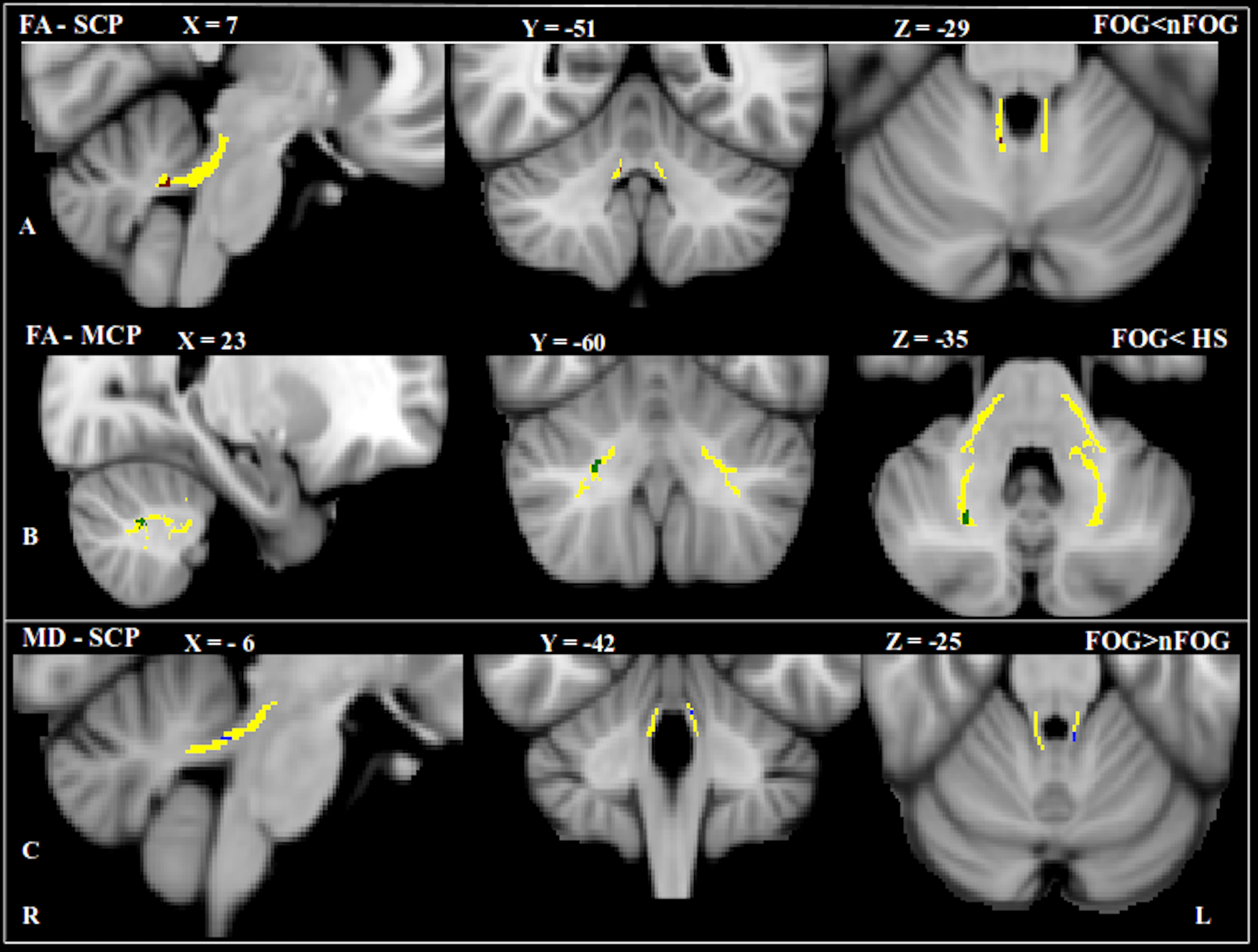
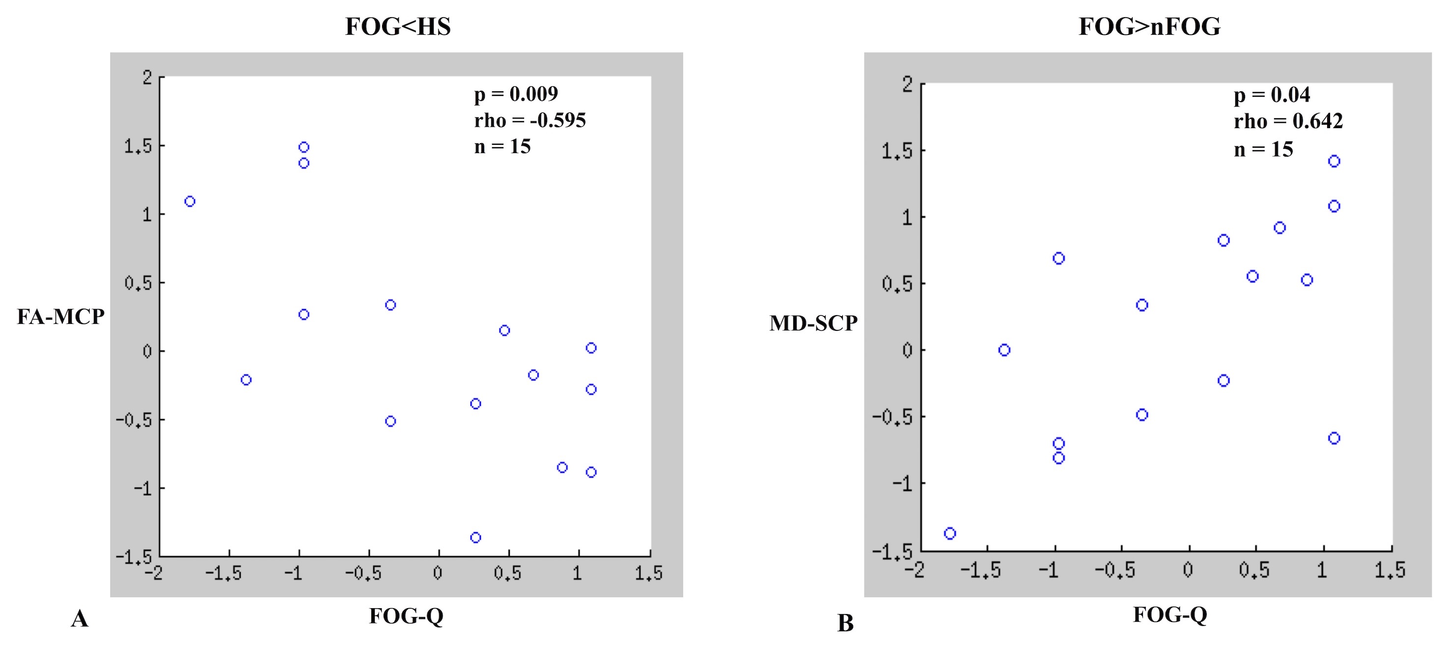
Results: PD-FOG patients showed lower FA in the Superior Cerebellar Peduncle (SCP) and the Middle Cerebellar Peduncle (MCP) than both PD-nFOG and HS [figure1A ][figure1B]. PD-FOG also showed higher MD in the Superior Cerebellar Peduncle (SCP) than PD-nFOG [figure1C]. FA at the Middle Cerebellar Peduncle (MCP) negatively correlated with FOG-Q in PD-FOG (p = 0.009; rho = – 0.595) [figure2A] and MD at the Superior Cerebellar Peduncle (SCP) positively correlated with FOG-Q in PD-FOG (p = 0.04; rho = 0.642) [figure2B].
Conclusions: Our results suggest the loss of fibers connecting the cerebellum with other brain structures in PD-FOG patients. Significant correlations support the role of the cerebellum in FOG pathogenesis, therefore underlines the importance of cerebellum with regards to locomotion.
References: 1. Nutt JG, Bloem BR, Giladi N, Hallett M, Horak FB, Nieuwboer A. Freezing of gait: moving forward on a mysterious clinical phenomenon. Lancet Neurol. 2011;10:734–44. 2. Youn J, Lee J-M, Kwon H, Kim JS, Son TO, Cho JW. Alterations of mean diffusivity of pedunculopontine nucleus pathway in Parkinson’s disease patients with freezing of gait. Parkinsonism Relat Disord. 2015;21:12–7.
To cite this abstract in AMA style:
K. Bharti, A. Suppa, S. Pietracupa, N. Upadhyay, C. Giannì, G. Leodori, F. Di Biaso, N. Modugno, N. Petasas, G. Grilla, A. Zampogna, A. Berardelli, P. Pantano. Structural Abnormalities in the Cerebellar Peduncles of patients with Freezing of Gait: A Diffusion Tensor Imaging Study [abstract]. Mov Disord. 2018; 33 (suppl 2). https://www.mdsabstracts.org/abstract/structural-abnormalities-in-the-cerebellar-peduncles-of-patients-with-freezing-of-gait-a-diffusion-tensor-imaging-study/. Accessed October 21, 2025.« Back to 2018 International Congress
MDS Abstracts - https://www.mdsabstracts.org/abstract/structural-abnormalities-in-the-cerebellar-peduncles-of-patients-with-freezing-of-gait-a-diffusion-tensor-imaging-study/


