Category: Neuroimaging (Non-PD)
Objective: To identify structural brain changes in patients with X-linked dystonia-parkinsonism (XDP) and non-manifesting carriers of the TAF1 SVA retrotransposon insertion (NMC).
Background: XDP is a neurodegenerative movement disorder characterized by severe adult-onset dystonia accompanied or followed by parkinsonism over the course of the disease. Post-mortem [1] and quantitative MR analyses [2,3] revealed marked striatal and pallidal atrophy in early disease stages already. Atrophy is pronounced and accompanied by iron deposition in the anterior (associative) putamen [4]. Advanced striatal decline in early disease stages is indicative of a long-lasting degenerative process preceding disease onset.
Method: T1-weighted cranial MRI scans were acquired at 1.5 Tesla in 10 male TAF1 NMCs, 25 matched controls negative for the disease-causing TAF1 mutation and 17 XDP patients. All investigations took place at the Makati Medical Center, Manila, Philippines. Voxel-based morphometry (VBM) was conducted using the CAT12 toolbox. Standardized video-taped neurological examinations were performed by two movement disorder specialists blinded to the genetic status.
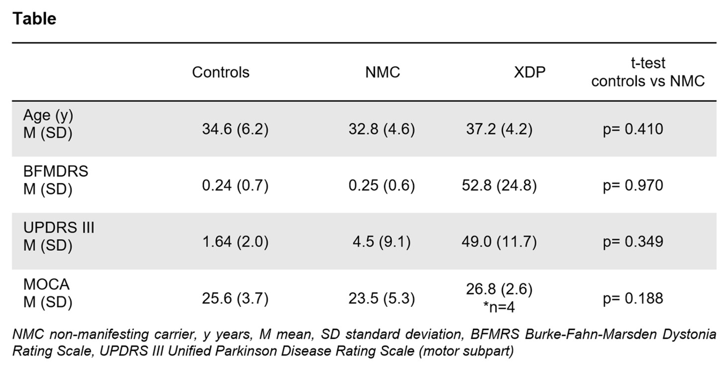
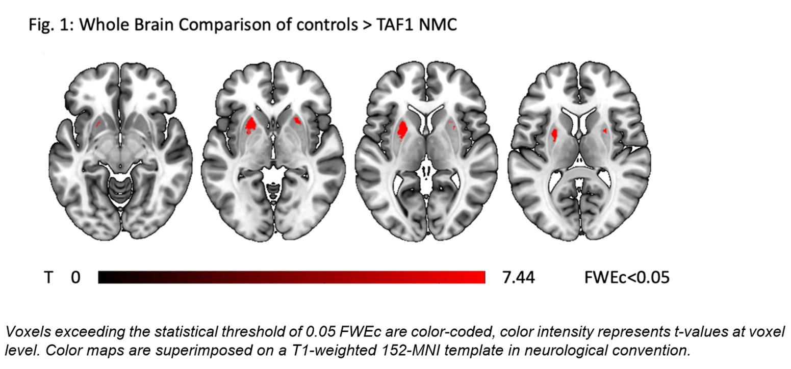
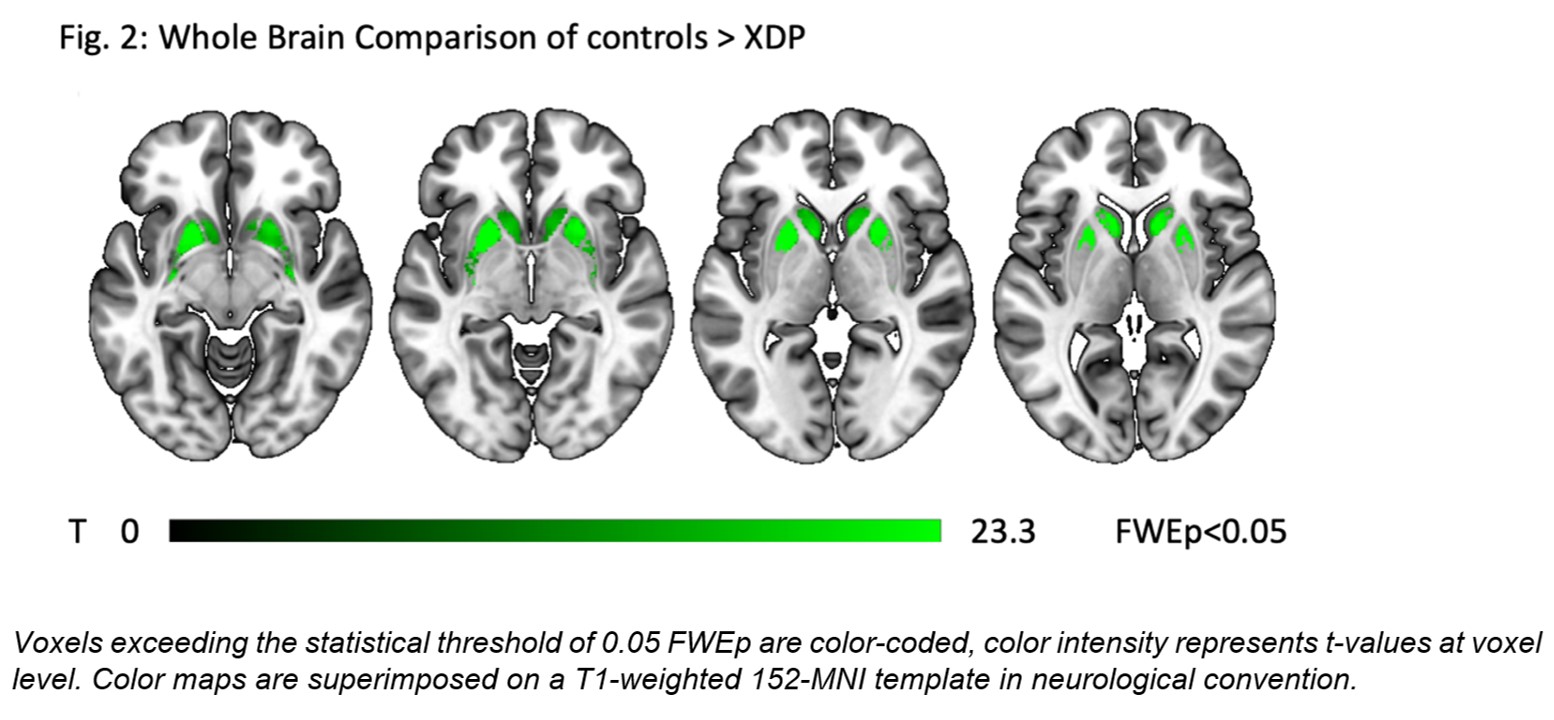
Results: XDP patients had a disease onset between the ages of 26 and 54 years, had a short disease duration of 0.5-6 years (2.5±1.6) and showed both, signs of dystonia and parkinsonism (table). Except for one asymptomatic individual with mild parkinsonian signs upon investigation, no unequivocal signs of dystonia or parkinsonism were present in NMCs (table). Whole-brain VBM revealed an isolated atrophy of the anterior putamen in NMC compared to controls (Fig. 1). XDP patients showed distinct gray matter loss in the putamen and caudate thereby confirming previous results (Fig. 2). Putaminal atrophy in NMCs was located exactly in the region of most severe atrophy in XDP patients.
[table1]
[figure 1]
[figure 2]
Conclusion: This study provides evidence for neurodegeneration in the presymptomatic phase of XDP. Isolated atrophy of the anterior putamen in NMCs indicates that the neurodegenerative process primarily involves the associate but not the sensorimotor part of the striatum. Thus, an impairment of executive functions may be present in the presymptomatic phase due the involvement of the fronto-striatal circuit. T1 imaging is a promising structural marker to identify TAF1 NMCs at risk to convert to XDP.
References: [1] Goto S, Lee LV, Munoz EL, et al. Functional anatomy of the basal ganglia in X-linked recessive Dystonia-parkinsonism. Ann Neurol 2005; 58:7–17 [2] Bruggemann N, Heldmann M, Klein C, et al. Neuroanatomical changes extend beyond striatal atrophy in X-linked dystonia parkinsonism. Parkinsonism Relat Disord 2016; 31:91–97 [3] Hanssen H, Heldmann M, Prasuhn J, et al. Basal ganglia and cerebellar pathology in X-linked dystonia-parkinsonism. Brain 2018; 141:2995–3008 [4] Hanssen H, Prasuhn J, Heldmann M, et al. Imaging gradual neurodegeneration in a basal ganglia model disease. Ann Neurol 2019; 86:517–526
To cite this abstract in AMA style:
H. Hanssen, M. Heldmann, J. Dy, J. Tantianpact, J. Steinhardt, A. Sprenger, A. Domingo, C. Reyes, R. Rosales, C. Klein, T. Münte, A. Westenberger, J. Oropilla, C. Diesta, N. Brüggemann. Striatal neurodegeneration in the presymptomatic phase of X-linked Dystonia-Parkinsonism [abstract]. Mov Disord. 2020; 35 (suppl 1). https://www.mdsabstracts.org/abstract/striatal-neurodegeneration-in-the-presymptomatic-phase-of-x-linked-dystonia-parkinsonism/. Accessed December 16, 2025.« Back to MDS Virtual Congress 2020
MDS Abstracts - https://www.mdsabstracts.org/abstract/striatal-neurodegeneration-in-the-presymptomatic-phase-of-x-linked-dystonia-parkinsonism/


