Category: Parkinson's Disease: Neuroimaging
Objective: Whether diffusion MRI (dMRI)-derived measures might help to understand sex-specific alterations of white-matter (WM) changes in Parkinson’s disease (PD) patients, with and without mild cognitive impairment (MCI), and healthy controls (HC) is unknown.
Background: PD presents itself with greater prevalence and age of onset in males as compared to females. However, none of the conventional single-tensor (ST) diffusion-MRI measures serve as a pathological marker to identify sex-specific differences in WM disorganization in PD. Also, whether beyond-single tensor dMRI-derived measures could serve as potential neuroimaging measure to identify sex-specific PD patients with MCI is unknown.
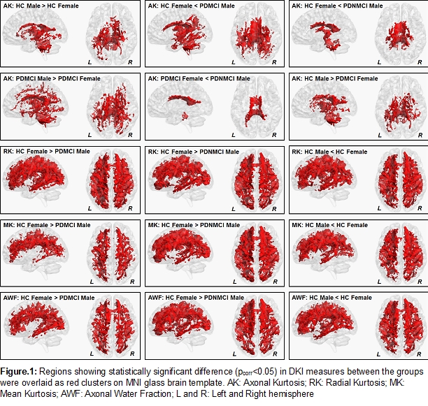
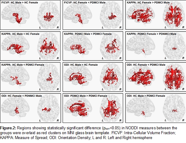
Method: 40 HC and 37 PD patients were recruited of which 22 PD patients were diagnosed with MCI based on results of a neuropsychological test battery. 71-directions dMRI was acquired at 3-shells (b=500s/mm2, 1000s/mm2, and 2500s/mm2) with a 3T Siemens scanner with an isotropic (1.5mm3) spatial resolution. Diffusion kurtosis (DKI) model and neurite orientation dispersion and density imaging (NODDI) model was fitted for every participant, and respective dMRI-derived measures were estimated in every brain voxel. WM skeleton was derived using tract-based-spatial-statistics. Each group was split into male and female before statistical comparisons. Nonparametric statistical comparisons for observing the difference in the means of beyond-single tensor dMRI-derived measures were conducted using permutation analysis of linear models with age, education, and handedness as nuisance regressors. Significance was established at family-wise-error-corrected (pcorr) <0.05.
Results: Handedness was different among the groups (p=0.02) (Table.1). Both DKI and NODDI revealed significant sex-specific differences between and within groups (Fig.1 and 2). Importantly, while both DKI and NODDI measures revealed sex-specific alterations in WM disorganization encompassing thalamo-cerebellar tracts in HC, PDMCI revealed sex-specific alterations in limbic, thalamic, and cerebellar tracts. Significant differences were also observed between sex and within sex of different groups, for both DKI and NODDI measures.
Conclusion: Multi-shell dMRI imaging is a promising avenue to explore sex-specific alterations in the WM microstructural integrity in PDMCI participants, and could be utilized to identify the most promising imaging biomarker which is sex-specific.
To cite this abstract in AMA style:
V. Mishra, K. Sreenivasan, D. Cordes, A. Ritter, Z. Mari, I. Litvan, J. Caldwell. Sex-specific white matter alterations in Parkinson’s disease with mild cognitive impairment [abstract]. Mov Disord. 2020; 35 (suppl 1). https://www.mdsabstracts.org/abstract/sex-specific-white-matter-alterations-in-parkinsons-disease-with-mild-cognitive-impairment/. Accessed April 21, 2025.« Back to MDS Virtual Congress 2020
MDS Abstracts - https://www.mdsabstracts.org/abstract/sex-specific-white-matter-alterations-in-parkinsons-disease-with-mild-cognitive-impairment/


