Session Information
Date: Wednesday, June 7, 2017
Session Title: Neuroimaging (Non-PD)
Session Time: 1:15pm-2:45pm
Location: Exhibit Hall C
Objective: To investigate the diagnostic value of semi-automated diagnostic algorithm using 3T-MR susceptibility-weighted image (SWI) for multiple system atrophy with parkinsonism (MSA-p).
Background: Iron deposition in the posterolateral putamen is an important feature which helps differentiate MSA-p from PD. Previous studies with MRI protocols which reflect iron content proved its usefulness as a diagnostic tool. However, most studies employed visual inspection. Even studies with quantitative methods had a problem of drawing the regions of interest (ROI) manually by the researcher, which is subject to rater-related inconsistency.
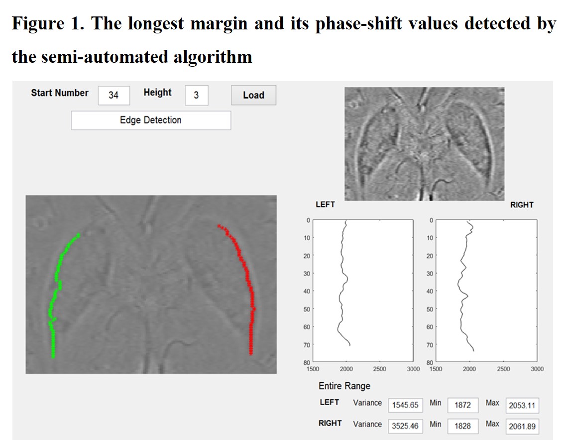
Methods: Our algorithm was developed by Step 1, detect the lateral margin of the putamen; Step 2, calculate the phase-shift values on the detected margin with varying numbers of pixels as ROI (Figure 1); Step 3, find the best differentiating ROI in terms of location and size. The receiver operating characteristic (ROC) curve analysis was used for investigating the most effective diagnostic condition. The algorithm was then applied to 26 MSA-p, 68 PD, and 41 normal control (NC) subjects with 3T-MR SWI.
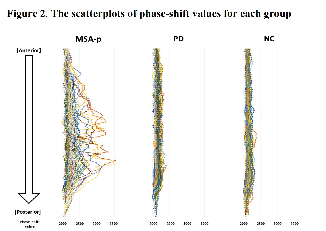
Results: The algorithm semi-automatically detected the lateral margin of the putamen in 125 (92.6%) of 135 subjects: 100% (26/26) in MSA-p, 88.2% (60/68) in PD, and 95.1% (39/41) in NC. The scatterplots in Figure 2 illustrate the raw phase-shift values along the lateral margin of the putamen for each group, which show an anterior to posterior gradient that is most marked in MSA-p. The representative value, modified from the phase-shift values within the posterior 4th portion of 5 sub-parts, was the best differentiating between MSA-p and PD with 80.8% of sensitivity, and 86.7% of sensitivity. (Our previous study using manual quantitation methods for phase-shift values had 77.8% of sensitivity and 76.0% of sensitivity. Reference 1)
Conclusions: Our MRI algorithm was able to detect lateral margin of the putamen semi-automatically, enabled us to calculate the phase-shift values in the best ROI, independent of a rater, and performed better in differentiating MSA-p from PD.
References: 1. Hwang I, Sohn CH, Kang KM, et al. Differentiation of Parkinsonism-Predominant Multiple System Atrophy from Idiopathic Parkinson Disease Using 3T Susceptibility-Weighted MR Imaging, Focusing on Putaminal Change and Lesion Asymmetry. AJNR American journal of neuroradiology 2015;36(12):2227-2234.
To cite this abstract in AMA style:
W.-W. Lee, H.-J. Kim, C.-H. Sohn, H. Park, B. Jeon. Semi-automated MRI algorithm using susceptibility weighted imaging for differentiating multiple system atrophy with parkinsonism from Parkinson Disease [abstract]. Mov Disord. 2017; 32 (suppl 2). https://www.mdsabstracts.org/abstract/semi-automated-mri-algorithm-using-susceptibility-weighted-imaging-for-differentiating-multiple-system-atrophy-with-parkinsonism-from-parkinson-disease/. Accessed February 12, 2026.« Back to 2017 International Congress
MDS Abstracts - https://www.mdsabstracts.org/abstract/semi-automated-mri-algorithm-using-susceptibility-weighted-imaging-for-differentiating-multiple-system-atrophy-with-parkinsonism-from-parkinson-disease/


