Objective: To investigate the mechanism by which retinoic acid-related orphan receptor alpha(RORα) regulates NAD+ levels to dopamine neuronal degeneration in Parkinson’s Disease (PD).
Background: PD is an age-related degenerative disease driven by neuroinflammation. RORα is a nuclear receptor involved in metabolic and inflammatory regulation, which has been found to be widely expressed in various cell types in central nervous system. NAD+ is a key signaling molecule in maintaining neuronal function and the concentration of NAD+ in the brain decreases with aging, but whether the NAD+ levels is associated with RORα in PD progression remains unknown.
Method: Blood samples of 25 PD patients and 25 age-matched healthy controls (HCs) were collected in EDTA-tubes from Wuhan Union Hospital. Peripheral blood mononuclear cell (PBMC) were isolated for subsequent qRT-PCR experiments. Preformed α-syn fibrils (PFFs) were utilized to induce α-syn overexpression in PD cell and mouse models. Motor function was evaluated. Neuronal degeneration markers were revealed by immunofluorescence and immunohistochemistry figures. NAD+ level was measured by ELISA kits. Single-cell RNA sequencing, bioinformatic analysis and ChIP experiments were used to determine the specific mechanism.
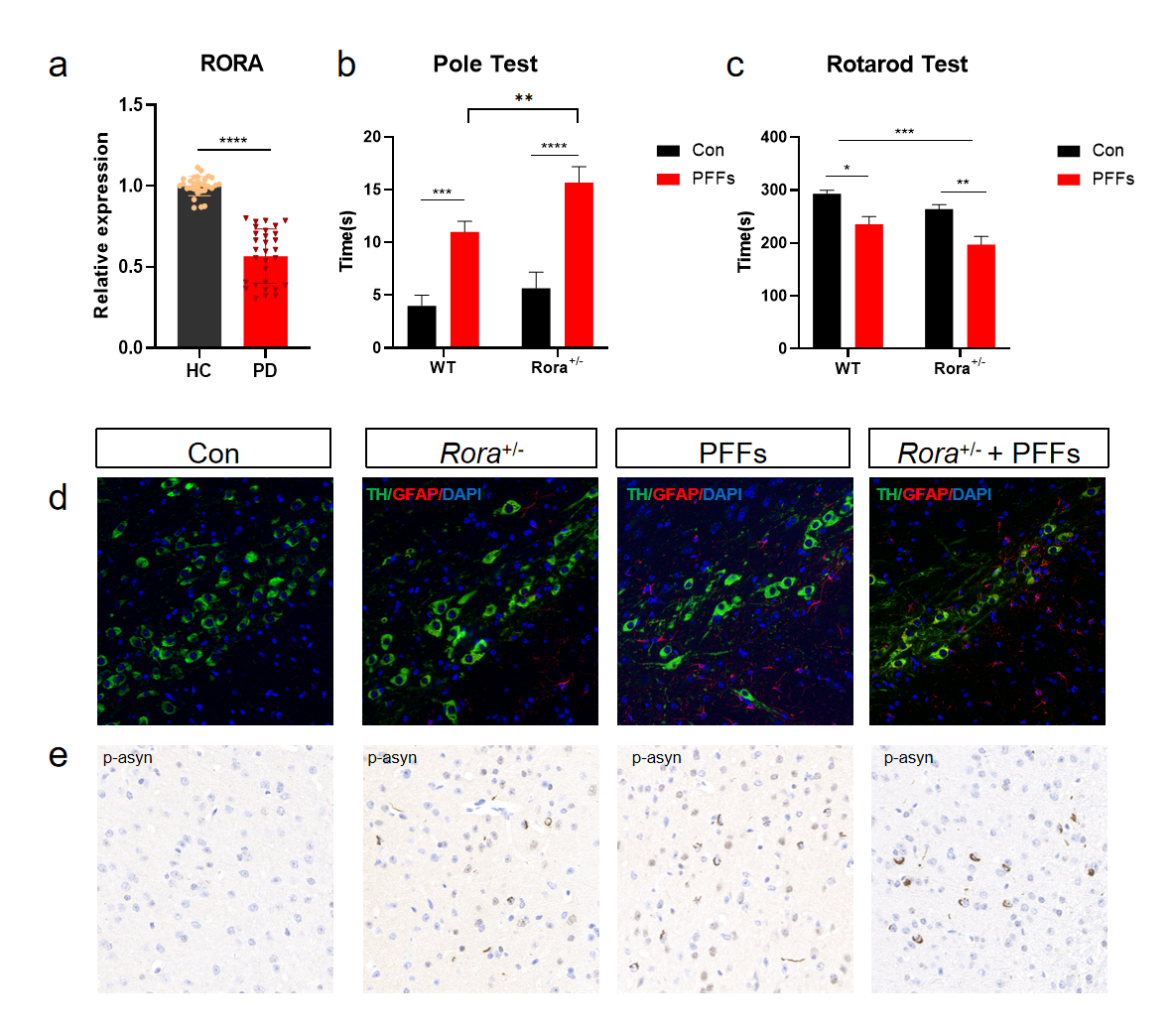
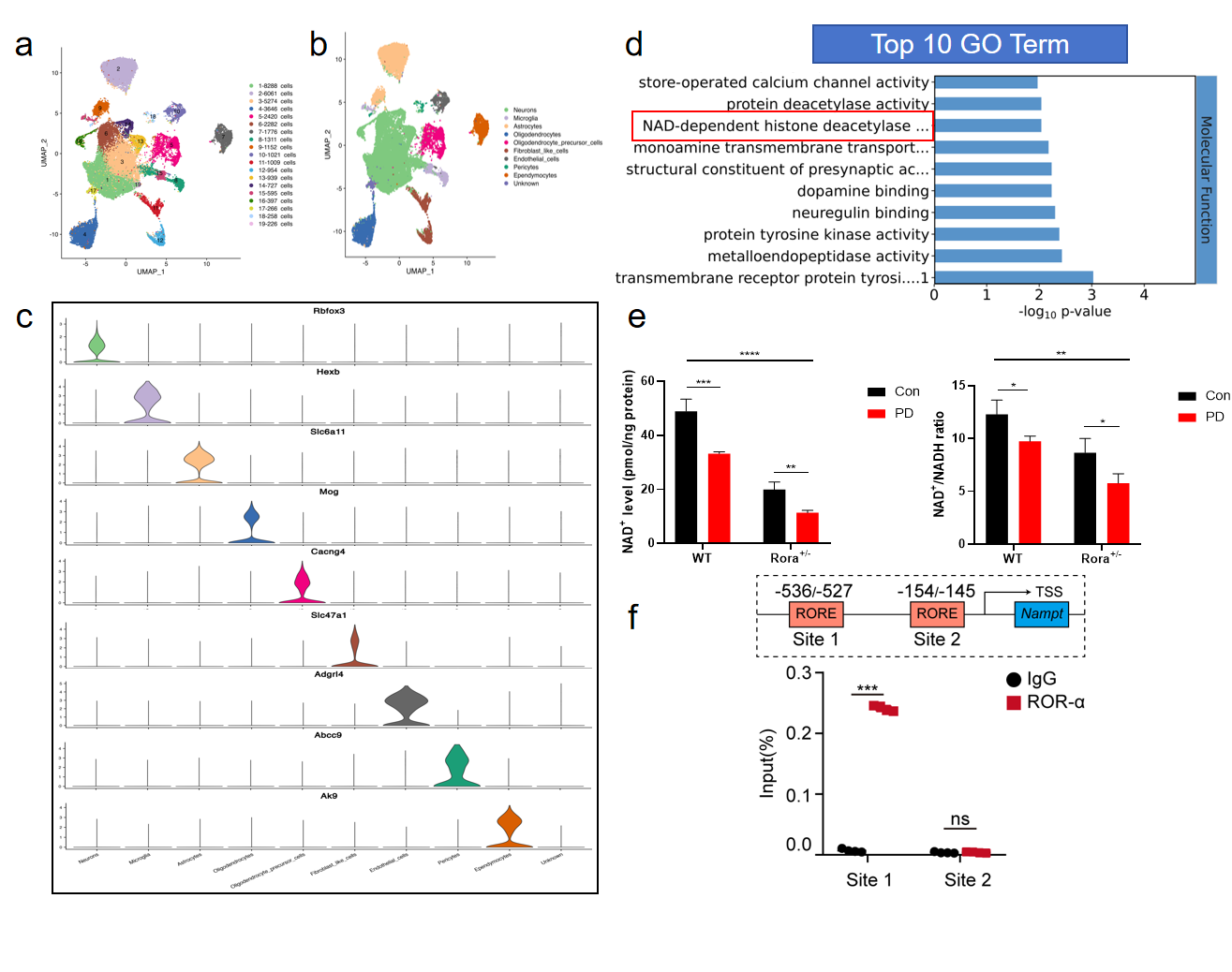
Results: We first identified that mRNA levels of RORα in PBMC of PD patients were down-regulated compared with healthy controls(Figure 1a). In vivo experiments suggested that Rora deficiency led to dopamine neurons loss, astrocytes activation, α-syn aggregation (especially increasing the phosphorylated-α-syn(S129) level) and finally motor function decrease(Figure 1b-e). We then did the single-cell RNA sequencing of mouse midbrain tissues and defined ten transcriptomic cell types(Figure 2a-c). For subtype identification, we focused most on the dopamine neurons. GO enrichment analysis showed the significant changes in NAD+ related biological process(Figure 2d). We examined the NAD+ of midbrain tissues in different groups and the results suggested NAD+ and NAD+/NADH ratio could be regulated by RORα in PD condition(Figure 2e). To confirm the regulatory effect of RORα on NAD+ level, we performed ChIP assay and the ChIP-qPCR showed that RORα could bind to NAMPT, the key enzyme of NAD+ synthesis (Figure 2f).
Conclusion: RORα regulated the transcription activity of the key enzyme NAMPT and then influence NAD+ level in the brain during PD progression.
RORα deficiency aggravated dopamine neurons loss
RORα reduced NAD+ levels via regulating NAMPT
References: Al-Zaid FS, Hurley MJ, Dexter DT, et al. Neuroprotective role for RORA in Parkinson’s disease revealed by analysis of post-mortem brain and a dopaminergic cell line. NPJ Parkinsons Dis. 2023;9(1):119.
Yang L, Shen J, Liu C, et al. Nicotine rebalances NAD+ homeostasis and improves aging-related symptoms in male mice by enhancing NAMPT activity. Nat Commun. 2023;14(1):900.
Li J, Liu H, Wang X, et al. Melatonin ameliorates Parkinson’s disease via regulating microglia polarization in a RORα-dependent pathway. NPJ Parkinsons Dis. 2022;8(1):90.
Schöndorf DC, Ivanyuk D, Baden P, et al. The NAD+ Precursor Nicotinamide Riboside Rescues Mitochondrial Defects and Neuronal Loss in iPSC and Fly Models of Parkinson’s Disease. Cell Rep. 2018;23(10):2976-2988.
To cite this abstract in AMA style:
JW. Li, HS. Liu, XY. Hu, T. Wang, N. Xiong. Rora deficiency aggravates dopamine neurons loss via reducing NAD+ level in Parkinson’s disease [abstract]. Mov Disord. 2024; 39 (suppl 1). https://www.mdsabstracts.org/abstract/rora-deficiency-aggravates-dopamine-neurons-loss-via-reducing-nad-level-in-parkinsons-disease/. Accessed April 20, 2025.« Back to 2024 International Congress
MDS Abstracts - https://www.mdsabstracts.org/abstract/rora-deficiency-aggravates-dopamine-neurons-loss-via-reducing-nad-level-in-parkinsons-disease/


