Category: Parkinson's Disease: Neuroimaging
Objective: To study the role of the recently identified direct subcortical cerebellum basal ganglia (C-BG) network in the pathophysiology of Parkinson’s Disease (PD).
Background: Even though the presence of direct subcortical connections between the cerebellum and basal ganglia in the form of disynaptic projections from the dentate nucleus to the contralateral Putamen and disynaptic projections from STN to the contralateral cerebellar cortex, has been established in non-human primates[1], their exact role in the pathophysiology of PD is yet to be fully understood.
Method: We recruited 20 healthy subjects (HS) (56.8 ± 8.7 yrs) with no history of neurological disorder and 20 Late PD patients (55.69 ± 9.96 yrs) based on disease duration and severity. They underwent MRI sessions (T1-w, Diffusion Weighted Imaging, and fMRI) in the ‘OFF’ stage (12-hour overnight withdrawal of drug). Also, the HS group underwent Addenbrook’s Cognitive Examination (ACE). The structural connections between direct subcortical C-BG networks were studied using Fixel-based analysis in the Mrtrix3[2] toolbox. Functional connectivity (FC) changes in the PD patients were studied using ROI-ROI analysis using the CONN toolbox using bilateral ROIs of the Putamen, Thalamus, Dentate, STN, Pontine nucleus, C7b, and Crus2.
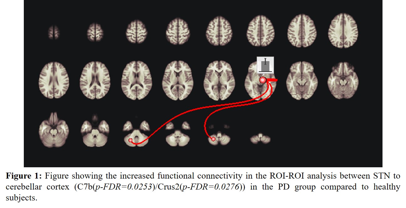
Results: Fixel-based analysis revealed, the fiber density (FD) metric of the cerebello-thalamo-putaminal tracts corelated with the registration/learning domain scores of ACE assessment (pcorrected=0.0117), while the FD metric of the subthalmo-cerebellar (Crus2) tracts corelated primarily with the memory domain (pcorrected=0.0431). The FC for the connections from STN-C7b (pFDR-corrected=0.0253) and STN-Crus2 (pFDR-corrected=0.0276) as in [figure1] were also found to increase in the PD group compared to HS.
Conclusion: The results reveal that subthalamo cerebellar tracts from STN-Crus2 are involved in memory processing while the dentato-thalamo-putaminal tracts are involved in learning/registration. Along with the FC results, this implies that one of the factors contributing to the pathophysiology of motor and non-motor functions in PD could be attributed to the abnormally increased FC in the recently identified pathway from the output of basal ganglia, the STN, to the cerebellar cortical region of C7b and Crus2, respectively.
References: [1] A. C. Bostan and P. L. Strick, “The basal ganglia and the cerebellum: nodes in an integrated network,” Nat Rev Neurosci, vol. 19, no. 6, pp. 338–350, Jun. 2018, doi: 10.1038/s41583-018-0002-7.
[2] J.-D. Tournier et al., “MRtrix3: A fast, flexible and open software framework for medical image processing and visualisation,” NeuroImage, vol. 202, p. 116137, Nov. 2019, doi: 10.1016/j.neuroimage.2019.116137.
To cite this abstract in AMA style:
V. Radhakrishnan, C. Gallea, S. Krishnan, B. Thomas, C. Kesavadas, A. Kishore. Role of the direct subthalamo-cerebellar tract in memory and learning: Implications for the pathophysiology of late-stage Parkinson’s Disease [abstract]. Mov Disord. 2023; 38 (suppl 1). https://www.mdsabstracts.org/abstract/role-of-the-direct-subthalamo-cerebellar-tract-in-memory-and-learning-implications-for-the-pathophysiology-of-late-stage-parkinsons-disease/. Accessed January 9, 2026.« Back to 2023 International Congress
MDS Abstracts - https://www.mdsabstracts.org/abstract/role-of-the-direct-subthalamo-cerebellar-tract-in-memory-and-learning-implications-for-the-pathophysiology-of-late-stage-parkinsons-disease/

