Objective: To identify EEG source level differences between patients with Parkinson’s disease (PD) and associated impulse control disorders (ICD) and patients without ICD.
Background: Previous research has identified a distinct neurophysiological profile in patients with PD and ICD. However, knowledge of the neurobiological substrate associated with these differences remains limited. In addition, the interaction between these brain regions and dopaminergic medications in these patients has not been thoroughly investigated.
Method: To investigate these regions, we performed resting state EEGs (3 minutes, eyes open) in 29 patients with ICD (ICD+) and 39 patients without ICD (ICD-). All recordings were made while the patients were on regular dopaminergic therapy (ON), of which EEG recordings were available from 14 ICD+ and 12 ICD- patients after overnight withdrawal of therapy (OFF). We performed spectral analysis on the EEG data and the sources of the primary frequency bands were estimated using the standardised low resolution electromagnetic tomography (sLORETA) solution. Comparative analyses between groups and dopaminergic treatment conditions were performed using Statistical Non-Parametric Mapping (SnPM toolbox).
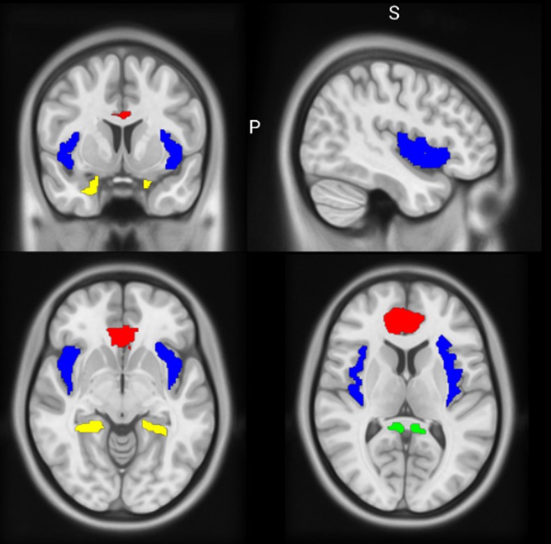
Results: Our results showed significant differences across the EEG frequency spectrum, in particular a marked increase in spectral power in the theta, alpha, beta 2 and beta 3 bands (Pperm < 0.05). Among these, a modulatory effect of dopaminergic therapy was observed only in the beta 3 band in ICD+ patients. Source analysis identified a group of brain regions responsible for variations in theta, alpha and beta 2 frequencies. These regions, including the insula, parahippocampal cortex and both anterior and posterior cingulate cortex, were found bilaterally (Pperm < 0.05) [figure 1]. Notably, the influence of dopaminergic treatment did not alter this specific set of regions.
Conclusion: Our findings suggest the presence of a specific neurobiological substrate that may explain the abnormal resting EEG patterns observed in patients with PD and ICD. Notably, this substrate appears to be unaffected by dopaminergic treatment. This finding may have important implications for understanding the neural mechanisms underlying these disorders and may inform future therapeutic strategies.
Figure 1.
To cite this abstract in AMA style:
E. Iglesias-Camacho, FJ. Gómez Campos, P. Franco Rosado, L. Garrote Espina, AM. Castellano-Gerrero, M. San Eufrasio, C. Perez-Calvo, L. Muñoz-Delgado, S. Jesús, D. Macías-García, E. Ojeda-Lepe, A. Adarmes-Gómez, F. Carrillo, JF. Martín-Rodriguez, P. Mir. Resting state EEG source analysis identifies distinct neural signature associated with impulse control disorders in PD [abstract]. Mov Disord. 2024; 39 (suppl 1). https://www.mdsabstracts.org/abstract/resting-state-eeg-source-analysis-identifies-distinct-neural-signature-associated-with-impulse-control-disorders-in-pd/. Accessed January 8, 2026.« Back to 2024 International Congress
MDS Abstracts - https://www.mdsabstracts.org/abstract/resting-state-eeg-source-analysis-identifies-distinct-neural-signature-associated-with-impulse-control-disorders-in-pd/

