Session Information
Date: Monday, October 8, 2018
Session Title: Parkinson's Disease: Neuroimaging And Neurophysiology
Session Time: 1:15pm-2:45pm
Location: Hall 3FG
Objective: Our aim was to investigate grey matter (GM) atrophy patterns in Parkinson’s disease patients with normal cognation (PD-NC) or with mild cognitive impairment(PD-MCI)using voxel based Morphometry (VBM), and to explore the relationship between GM volumes of the whole brain and globe cognition, and to reveal between GM atrophic regions of and cognitive performance in specific domains.
Background: Previous studies can suggest us that cognitive function may decline gradually with expansion of brain regions of grey matter atrophy in PD patients[1,2]. However, the correlation between atrophic brain regions and performance in specific cognitive domains in PD patients is not clear.
Methods: Seventy-two non-demented PD patients and 12 healthy controls (HC) underwent comprehensive clinical and neuropsychological assessment and 3.0T magnetic resonance scanning. VBM on structural MRI was applied to explore atrophic regions of GM between HC, PD-NC and PD-MCI groups. Regression analyses were performed to evaluate relationships between atrophic regions and performance in five specific cognitive domains (memory, executive function, language, visuospatial function and attention/working memory).
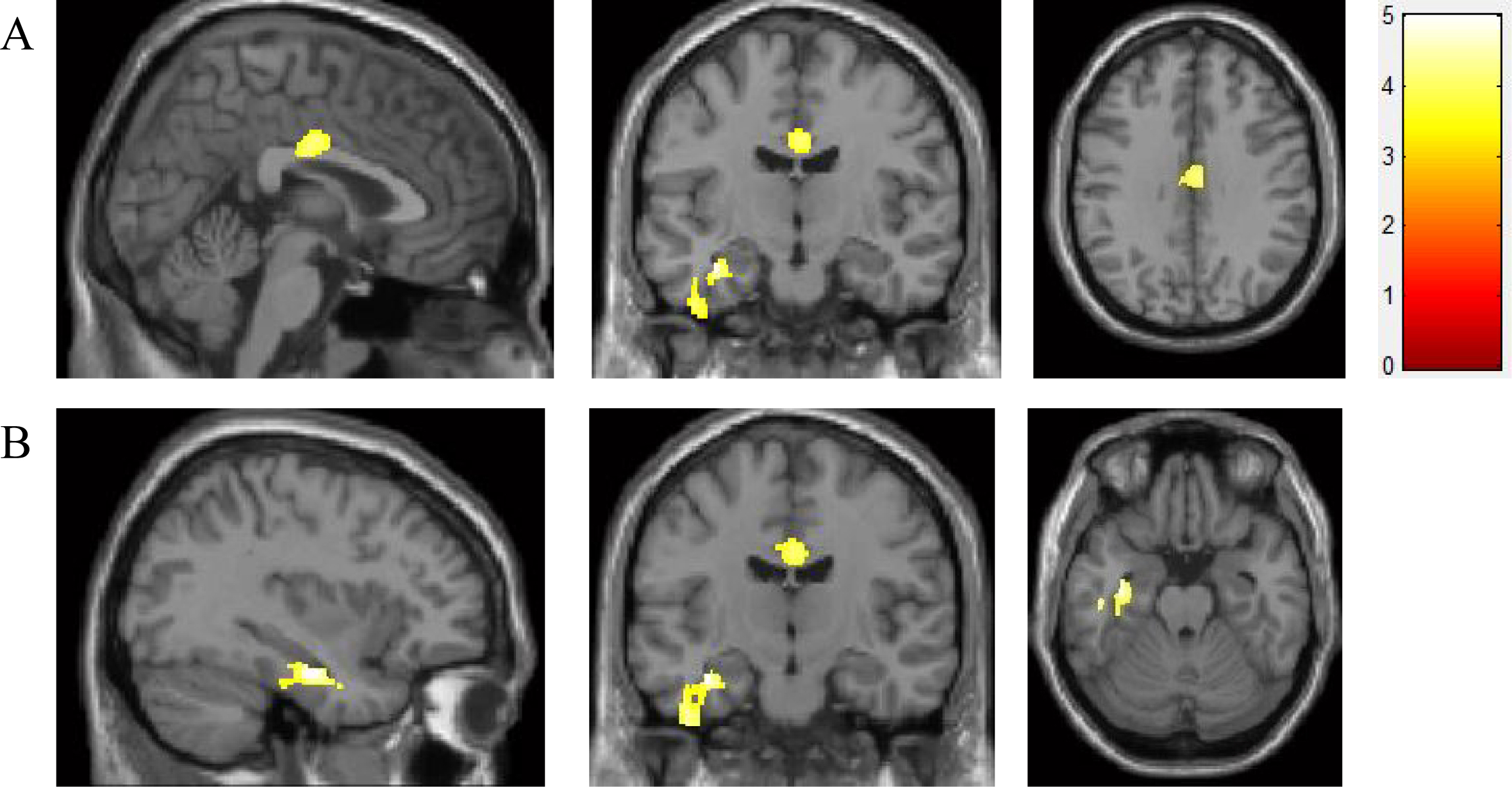
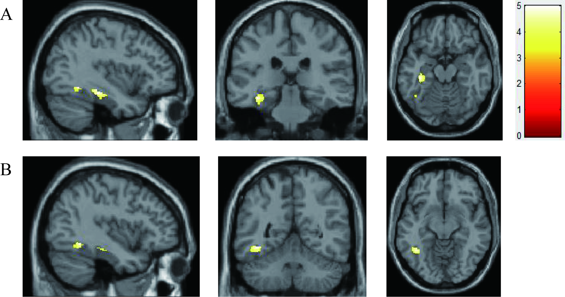
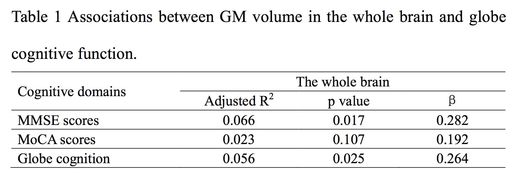
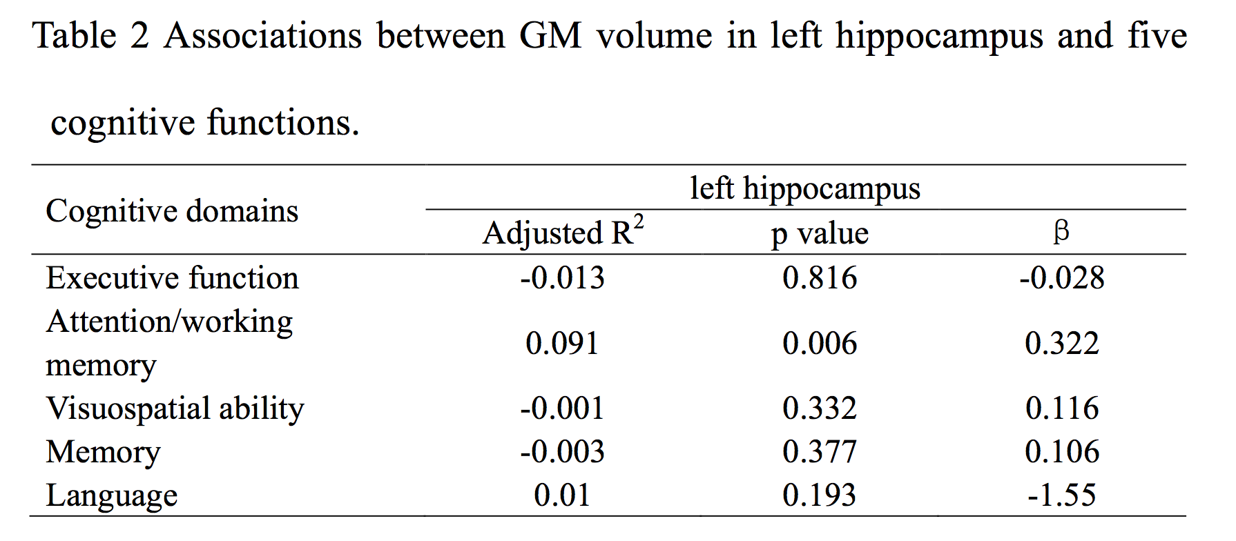
Results: Compared to HC, atrophic regions in PD-NC and PD-MCI patients were showed in the right middle cingulum, left middle occipital, and left superior occipital(Figure1 and 2). Moreover, compared to PD-NC, cortical atrophy in PD-MCI patients was found in the left fusiform gyrus, left hippocampus, inferior temporal gyrus(Figure 3). GM volumes of the whole brain were associated with globe cognition(Table 1). Attention/working memory was associated left hippocampus, suggesting a relationship between cognitive impairment and atrophy of left hippocampus in the whole PD sample(Table 2).
Conclusions: PD-MCI is closely associated with atrophy of GM in the left inferior temporal gyrus, left fusiform and left hippocampus. The loss of GM volume may serve as a marker for diagnosing initial cognitive decline in PD.
References: [1]. Noh SW, Han YH, Mun CW, et al. Analysis among cognitive profiles and gray matter volume in newly diagnosed Parkinson’s disease with mild cognitive impairment. J Neurol Sci. 2014 347: 210-213. [2]. Melzer TR, Watts R, MacAskill MR, et al. Grey matter atrophy in cognitively impaired Parkinson’s disease. Journal of Neurology, Neurosurgery & Psychiatry. 2012 83: 188-194.
To cite this abstract in AMA style:
K. Nie, M. Mei, Y. Gao, M. Guo, B. Huang, Z. Huang, L. Wang, J. Zhao, Y. Zhang, D. Xiong, L. Wang. Relationships between specific subtypes of mild cognitive impairment and grey matter atrophy in Parkinson’s disease [abstract]. Mov Disord. 2018; 33 (suppl 2). https://www.mdsabstracts.org/abstract/relationships-between-specific-subtypes-of-mild-cognitive-impairment-and-grey-matter-atrophy-in-parkinsons-disease/. Accessed December 17, 2025.« Back to 2018 International Congress
MDS Abstracts - https://www.mdsabstracts.org/abstract/relationships-between-specific-subtypes-of-mild-cognitive-impairment-and-grey-matter-atrophy-in-parkinsons-disease/


