Category: Parkinson's Disease: Neuroimaging
Objective: To examine resting-state functional Magnetic Resonance Imaging in a group of Parkinson’s Disease patients, used to characterize changes in functional connectivity of the olfactory network.
Background: Resting state functional MRI (rs-fMRI), measures spontaneous low-frequency fluctuations in blood (BOLD) signal in subjects at rest. Several studies are exploring the use of rs-fMRI techniques in examining possible functional dysconnectivity effects in Parkinson’s Disease (PD). Paralleling the neuropathological progression of PD, rs-fMRI changes (both increase and decrease functional connectivity) involving the dopaminergic cortico-striatal and mesolimbic-striatal loops have been consistently detected in PD patients.
Hyposmia is a well-established and early non-motor symptom of PD. Research has suggested its role as a potential biomarker of PD progression and cognitive decline.
Despite the advances in both olfactory and resting-state fMRI, it is still not fully understood how olfactory dysfunction in PD is reflected in the activation pattern of the olfactory network and results from previous olfactory fMRI studies have been inconsistent.
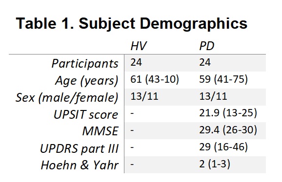
Method: -Subjects: 24 PD patients (PDs) with mild-moderate bilateral disease in the OFF dopaminergic state and 30 matched healthy volunteers (HVs) were scanned (Table 1).
-Imaging:
Scans collected on 3T scanners (GE Signa HDx)
T1 -weighted anatomical images (for localization)
T2-weighted echo planar images were collected during rest with eyes closed for 6 minutes
-Preprocessing: rs-fMRI data were pre-processed using the AFNI scripts afni_proc.py
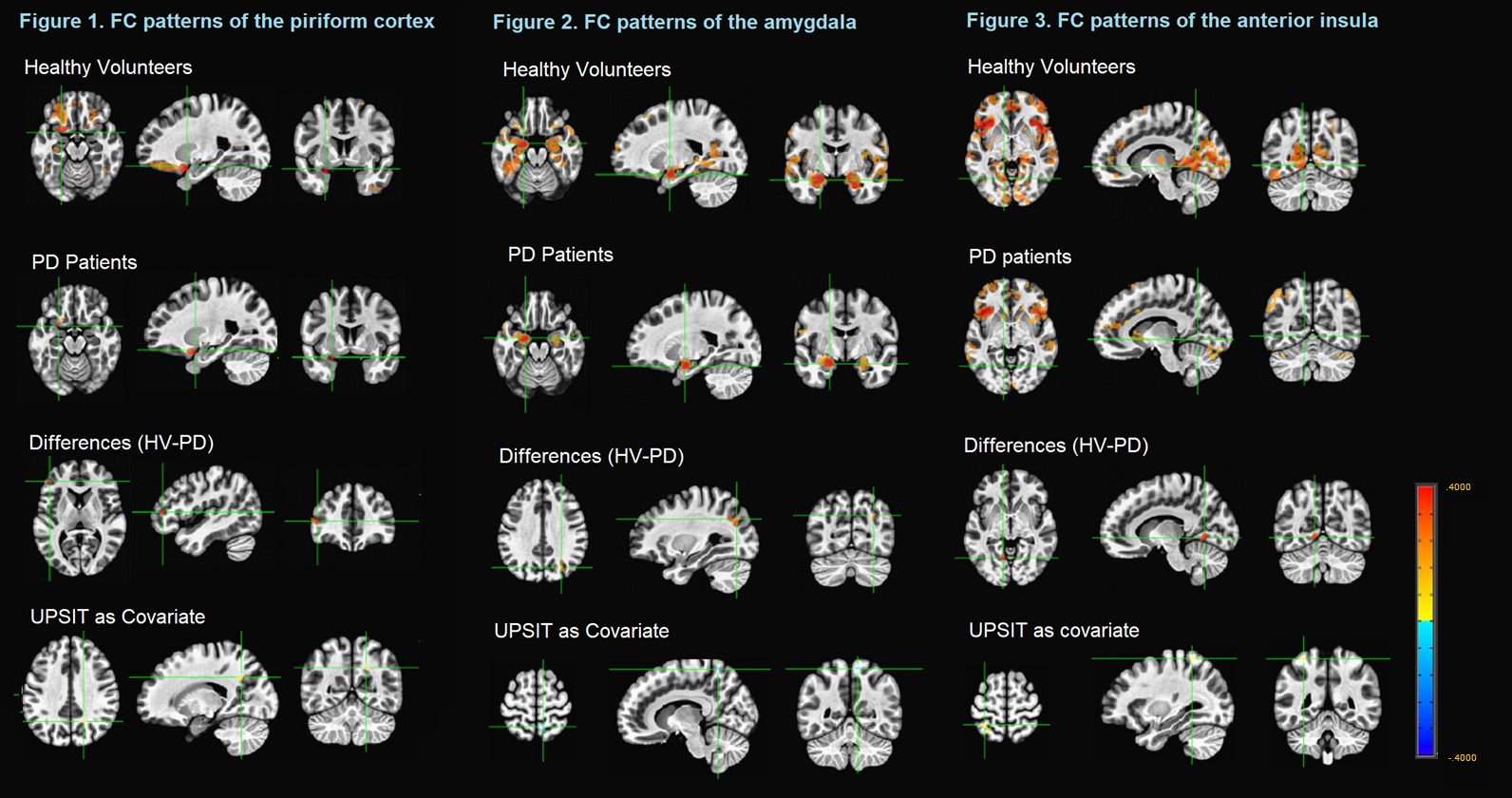
-Seed-based Correlation Analysis: Functional connectivity (FC) analyses were conducted using 3dGroupInCorr in AFNI to analyze key Regions of Interest (ROIs) in the olfactory network. The fronto-parietal ROI time-series were computed by selecting a seed location based on predefined coordinates. Key olfactory areas, 3 regions receiving direct olfactory bulbar input, including the piriform cortex, amygdala and anterior insula which have been reported to be important components of the olfactory subnetworks
Results: As shown in Figure 1-3
Conclusion: There are differences in the FC between HV and PDs when analyzing ROIs) in the olfactory network.
Changes in the sense of smell (as found by the UPSIT score), showed changes in the FC in Higher-order integrative cortical areas.
References: 1. Arnold TC, You Y, Ding M, Zuo XN, de Araujo I, Li W. Functional Connectome Analyses Reveal the Human Olfactory Network Organization. eNeuro. 2020 Aug 10;7(4):ENEURO.0551-19.2020. 2. Georgiopoulos C, Witt ST, Haller S, Dizdar N, Zachrisson H, Engström M, Larsson EM. A study of neural activity and functional connectivity within the olfactory brain network in Parkinson’s disease. Neuroimage Clin. 2019;23:101946.
To cite this abstract in AMA style:
L. Pesantez Pacheco, S. Horovitz. Relationship Between Hyposmia and Olfactory Network Connectivity in Patients With Parkinson’s Disease [abstract]. Mov Disord. 2021; 36 (suppl 1). https://www.mdsabstracts.org/abstract/relationship-between-hyposmia-and-olfactory-network-connectivity-in-patients-with-parkinsons-disease/. Accessed April 22, 2025.« Back to MDS Virtual Congress 2021
MDS Abstracts - https://www.mdsabstracts.org/abstract/relationship-between-hyposmia-and-olfactory-network-connectivity-in-patients-with-parkinsons-disease/


