Category: Parkinson's Disease: Neuroimaging
Objective: Assess to what extent free-water(FW) as measured by diffusion-weighted MRI(dMRI) is a neuroimaging marker of glial activation, neuroinflammation, and neurodegeneration in de novo Parkinson’s disease(PD) patients from the Parkinson’s Progression Markers Initiative(PPMI).
Background: Recent studies reported that the progressive FW accumulation particularly in the substantia nigra(SN) was linked to worsening of Parkinson’s symptoms, suggesting FW as a non-invasive imaging marker to track the progressive degeneration of neurons in PD1 with potential as a clinical trial outcome measure.Though, the underlying mechanism of changes in FW levels in PD has yet to be understood.We evaluated how cerebrospinal fluid(CSF) markers of glial activation, neuroinflammation, and neurodegeneration relates to FW in de novo PD patients from PPMI.
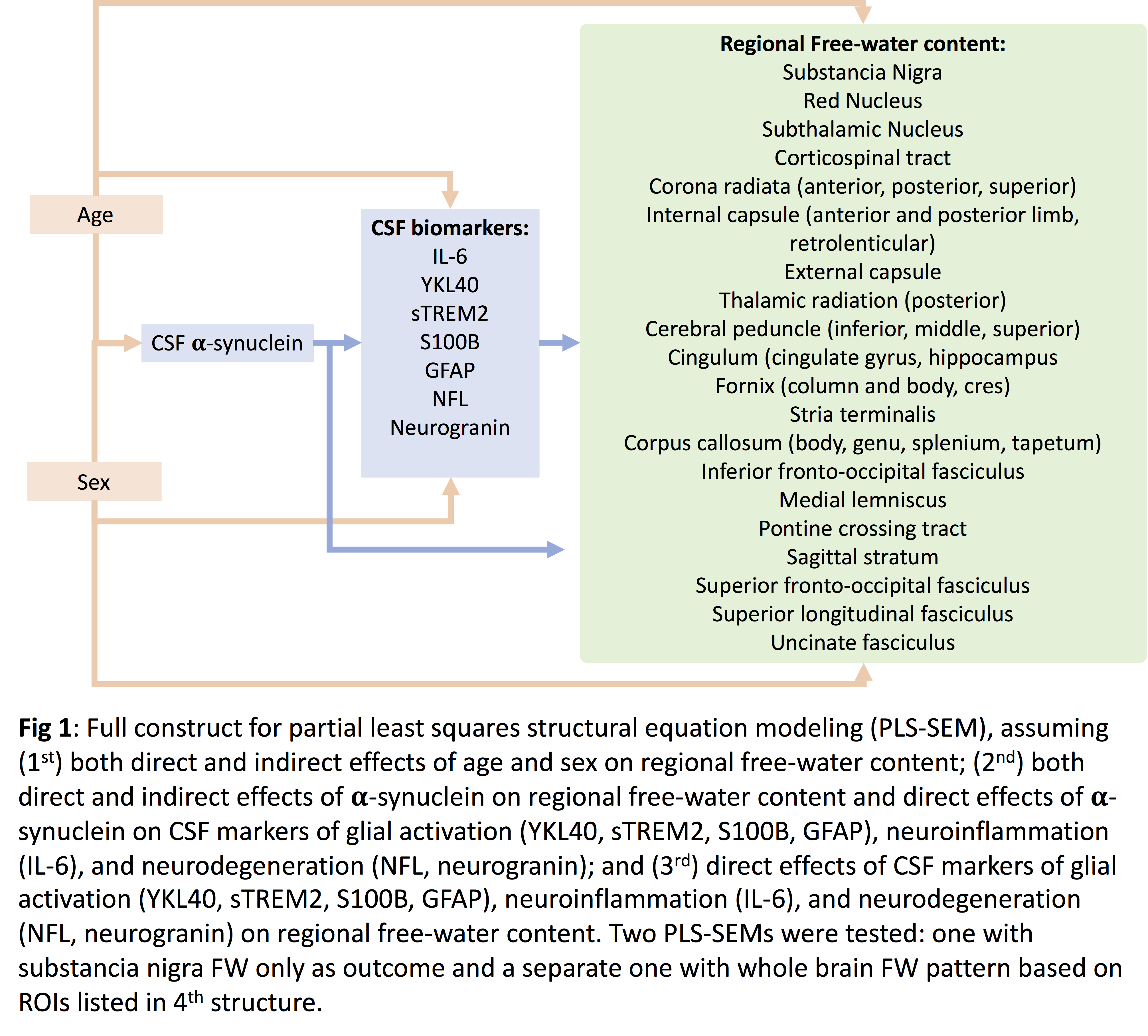
Method: 17 PD patients with valid baseline dMRI data and CSF Roche Elecsys Neuro Toolkit Immunoassays were analyzed.Each dMRI acquisition was non-linearly registered in MNI space using the ANTs software package.The white matter ROIs and subcortical nuclei were defined using JHU-MNI atlas.The diffusion tensor was calculated using a bi-tensor model, providing an estimate of FW2.CSF markers of glial activity(GFAP, sTREM2, S100b, and YKL40), inflammation(IL-6), synaptic dysfunction(neurogranin), neuroaxonal damage(NFL), and alpha-syn were tested.Partial least squares structural equation modeling (PLS-SEM) was used for assessing direct and indirect effects of observed and latent variables on regional FW levels3.Full construct for PLS-SEM is shown in Fig 1.
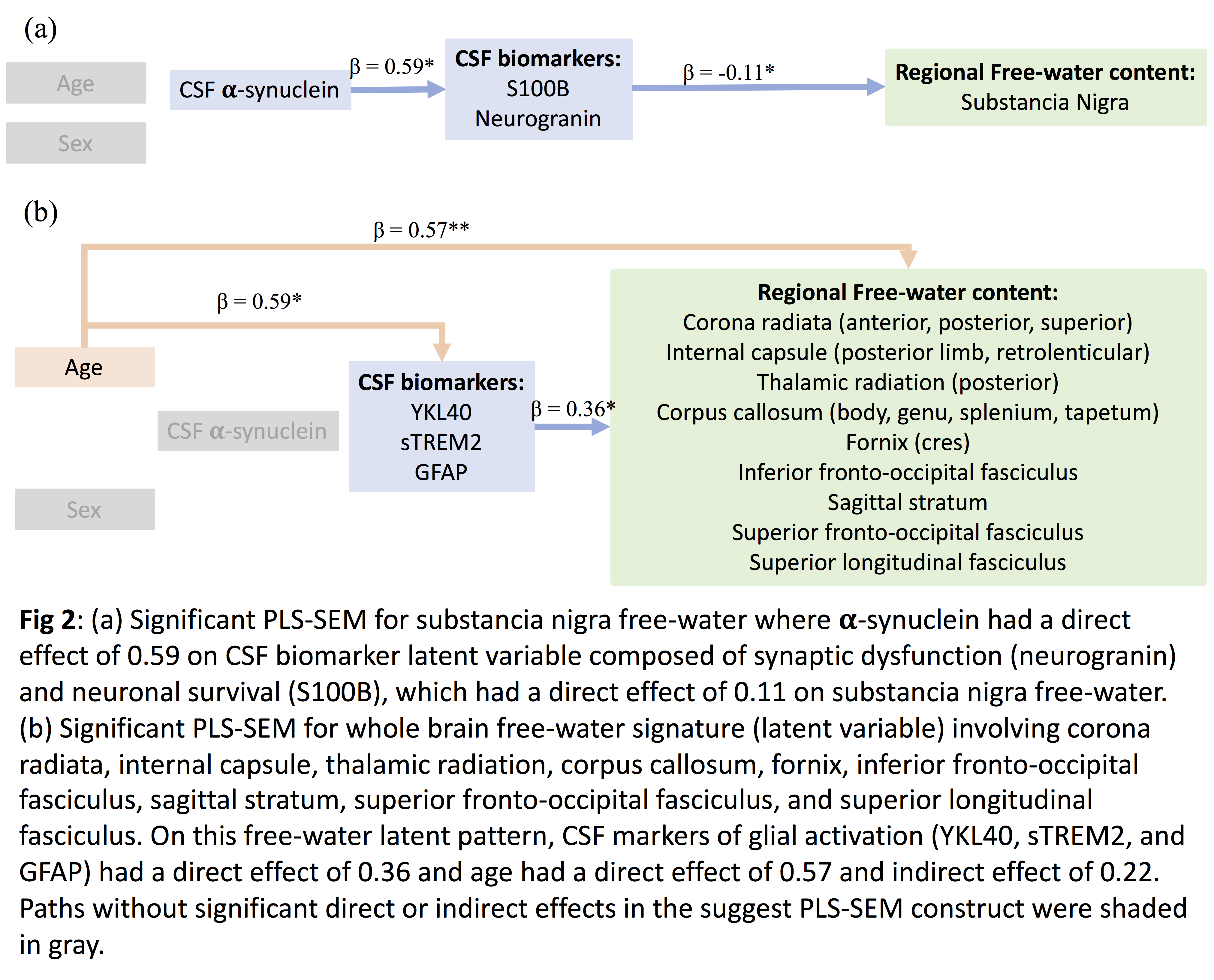
Results: Structured SN FW only and whole brain FW associations were shown in Fig 2.
Conclusion: Variance in SN FW was associated with synaptic dysfunction and neuronal survival(CSF neurogranin and S100b) biomarkers that were associated with higher alpha-synuclein levels. In contrast, variance in FW in wide-spread WM regions(corona radiata, internal capsule, thalamic radiation, corpus callosum, fronto-occipital fasciculus, superior longitudinal fasciculus, fornix, and sagittal stratum) was associated with CSF glial activation biomarkers(sTREM2, YKL40, GFAP) and advanced age.These differential associations with glial activation and neurodegeneration markers support regional FW as a promising stage-specific progression biomarker in PD that could help identify protective therapies in clinical trials.
References: 1 Burciu, R. G. et al. Progression marker of Parkinson’s disease: a 4-year multi-site imaging study. Brain 140, 2183-2192, doi:10.1093/brain/awx146 (2017). 2 Pasternak, O., Sochen, N., Gur, Y., Intrator, N. & Assaf, Y. Free water elimination and mapping from diffusion MRI. Magn Reson Med 62, 717-730, doi:10.1002/mrm.22055 (2009). 3 Hair Joseph, F. When to use and how to report the results of PLS-SEM. European Business Review 31, 2-24, doi:10.1108/EBR-11-2018-0203 (2019).
To cite this abstract in AMA style:
D. Tosun, R. Ellis. Regional free-water differentially associated with glial activation, neuroinflammation, and neurodegeneration in de novo Parkinson’s disease [abstract]. Mov Disord. 2020; 35 (suppl 1). https://www.mdsabstracts.org/abstract/regional-free-water-differentially-associated-with-glial-activation-neuroinflammation-and-neurodegeneration-in-de-novo-parkinsons-disease/. Accessed December 29, 2025.« Back to MDS Virtual Congress 2020
MDS Abstracts - https://www.mdsabstracts.org/abstract/regional-free-water-differentially-associated-with-glial-activation-neuroinflammation-and-neurodegeneration-in-de-novo-parkinsons-disease/


