Category: Parkinson's Disease: Neuroimaging
Objective: To shed light on the genes underlying increased cortical iron deposition in Parkinson’s disease (PD).
Background: Oxidative stress secondary to brain iron accumulation is a potential pathomechanism for neurodegeneration in PD [1]. Brain iron can be imaged using quantitative susceptibly mapping (QSM) [2]. We identify the pattern of healthy brain gene transcription underlying increased brain iron in PD.
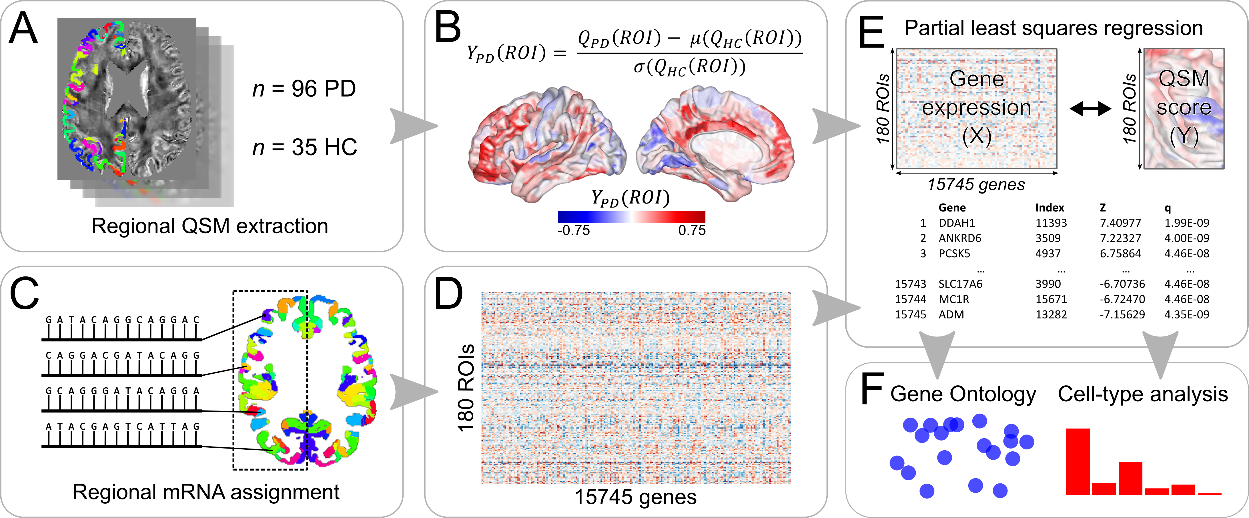
Method: We obtained susceptibility-weighted MRI images for 96 PD patients and 35 controls using a spoiled-GRE sequence. Phase images were unwrapped with the Laplacian method and brain masks calculated using the BET2 algorithm. Phase pre-processing was completed using Laplacian boundary value extraction and variable spherical mean-value filtering. Susceptibility maps were estimated using Multi-Scale Dipole Inversion[3]. Using advanced normalisation tools (ANTs), a study-wise template was created from native space T1 images. QSM images and 180 left-cortical regions of the Glasser atlas[4] were transformed into this space using ANTs. PD age- and sex-adjusted regional means were normalised to the control mean by a Z-score transformation. Microarray data from six donors were obtained from the Allen Institute for Brain Science[5]. Each tissue sample was assigned to one of the 180 regions and expression levels for each gene were compiled to form a 180×15745 regional transcription matrix[6]. We used partial least squares regression to investigate associations between the healthy brain transcriptome and QSM differences in PD. We used g:Profiler[7] for gene ontological (GO) analysis of the most strongly weighted genes. We filtered the resulting list of GO terms, retaining only those significantly enriched at corrected P<0.05. We investigated the cell types these genes were predominantly expressed in using expression weighted cell type analysis[8] [figure1].
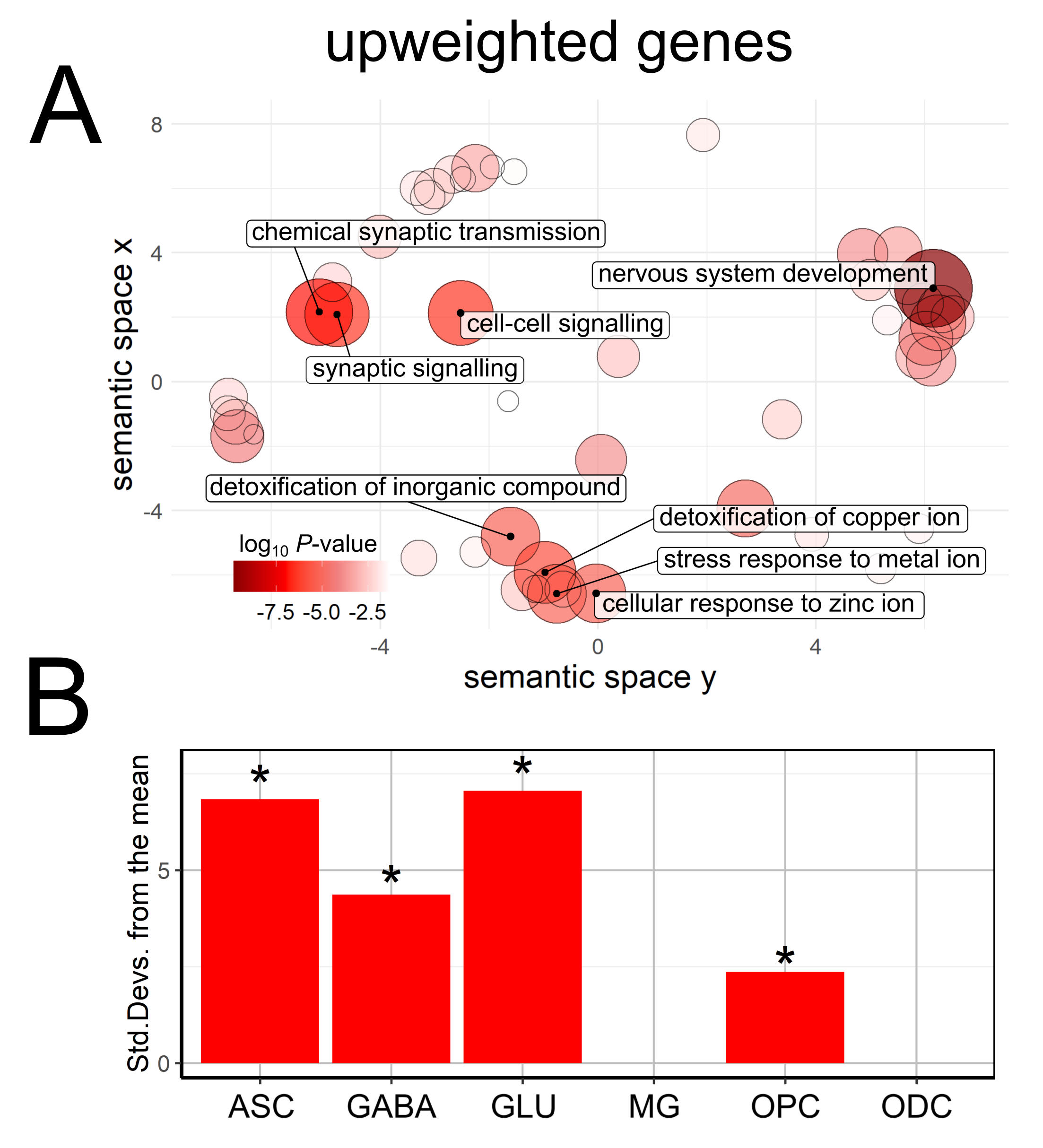
Results: Increased cortical magnetic susceptibility, likely reflecting iron accumulation in PD, was associated with higher intrinsic expression of genes involved in metal detoxification and synaptic function. Regional differences in the probable proportion of astrocytes, neurons and oligodendrocyte precursor cells were also found [figure2].
Conclusion: Our findings suggest that regional gene expression related to metal detoxification and synaptic function may contribute to selective vulnerability driving neurodegeneration in PD.
References: 1. Ward RJ, Zucca FA, Duyn JH, et al. The role of iron in brain ageing and neurodegenerative disorders. Lancet Neurol. 2014;13(10):1045–1060. 2. Shmueli K, De Zwart JA, Van Gelderen P, et al. Magnetic susceptibility mapping of brain tissue in vivo using MRI phase data. Magn. Reson. Med. 2009;62(6):1510–1522. 3. Acosta-Cabronero J, Milovic C, Mattern H, et al. A robust multi-scale approach to quantitative susceptibility mapping. Neuroimage 2018;183(February):7–24. 4. Glasser MF, Coalson TS, Robinson EC, et al. A multi-modal parcellation of human cerebral cortex. Nature 2016;536(7615):171–178. 5. Hawrylycz MJ, Lein ES, Guillozet-Bongaarts AL, et al. An anatomically comprehensive atlas of the adult human brain transcriptome. Nature 2012;489(7416):391–399. 6. Arnatkevic̆iūtė A, Fulcher BD, Fornito A. A practical guide to linking brain-wide gene expression and neuroimaging data. Neuroimage 2019;189:353–367. 7. Raudvere U, Kolberg L, Kuzmin I, et al. g:Profiler: a web server for functional enrichment analysis and conversions of gene lists (2019 update). Nucleic Acids Res. 2019;47:191–198. 8. Skene NG, Grant SGN. Identification of vulnerable cell types in major brain disorders using single cell transcriptomes and expression weighted cell type enrichment. Front. Neurosci. 2016;10(JAN).
To cite this abstract in AMA style:
G. Thomas, A. Zarkali, M. Ryten, K. Shmueli, AL. Gil Martinez, LA. Leyland, P. Mccolgan, J. Acosta-Cabronero, A. Lees, R. Weil. Regional brain iron and gene expression variation in Parkinson’s disease [abstract]. Mov Disord. 2021; 36 (suppl 1). https://www.mdsabstracts.org/abstract/regional-brain-iron-and-gene-expression-variation-in-parkinsons-disease/. Accessed April 19, 2025.« Back to MDS Virtual Congress 2021
MDS Abstracts - https://www.mdsabstracts.org/abstract/regional-brain-iron-and-gene-expression-variation-in-parkinsons-disease/


