Session Information
Date: Tuesday, June 6, 2017
Session Title: Rare Genetic and Metabolic Diseases
Session Time: 1:45pm-3:15pm
Location: Exhibit Hall C
Objective: To better understand striatal pathology in X-linked Dystonia-Parkinsonism (XDP) by combining T1 and Magnetization Transfer (MT) Imaging.
Background: XDP is a neurodegenerative disorder characterized by severe adult-onset dystonia followed by parkinsonism over the course of the disease. Post-mortem and quantitative MR analyses revealed severe striatal and pallidal atrophy even in early disease stages. MT imaging is a sensitive method to reveal microstructural alterations of tissue integrity and may be helpful to differentiate between demyelination (very low MT ratio (MTR)) and edema (slightly reduced MTR).
Methods: T1-weighted and MT images were acquired from 10 male XDP-patients (age: M= 42.6, SD= 7.6; mean disease duration: 3.4 years, range: 1-6 years) and 16 ethnicity-matched male healthy controls (age: 35.6, 7.3) at 3 Tesla.
To detect a pattern or gradient of neurodegeneration, three regions of interest (ROI) were defined (rostral, central and caudal putamen), reflecting functionally divergent regions of the putamen. Mean voxel intensity and MTR values were extracted with FSL. To test for statistical differences a mixed model ANOVA using SPSS was performed.
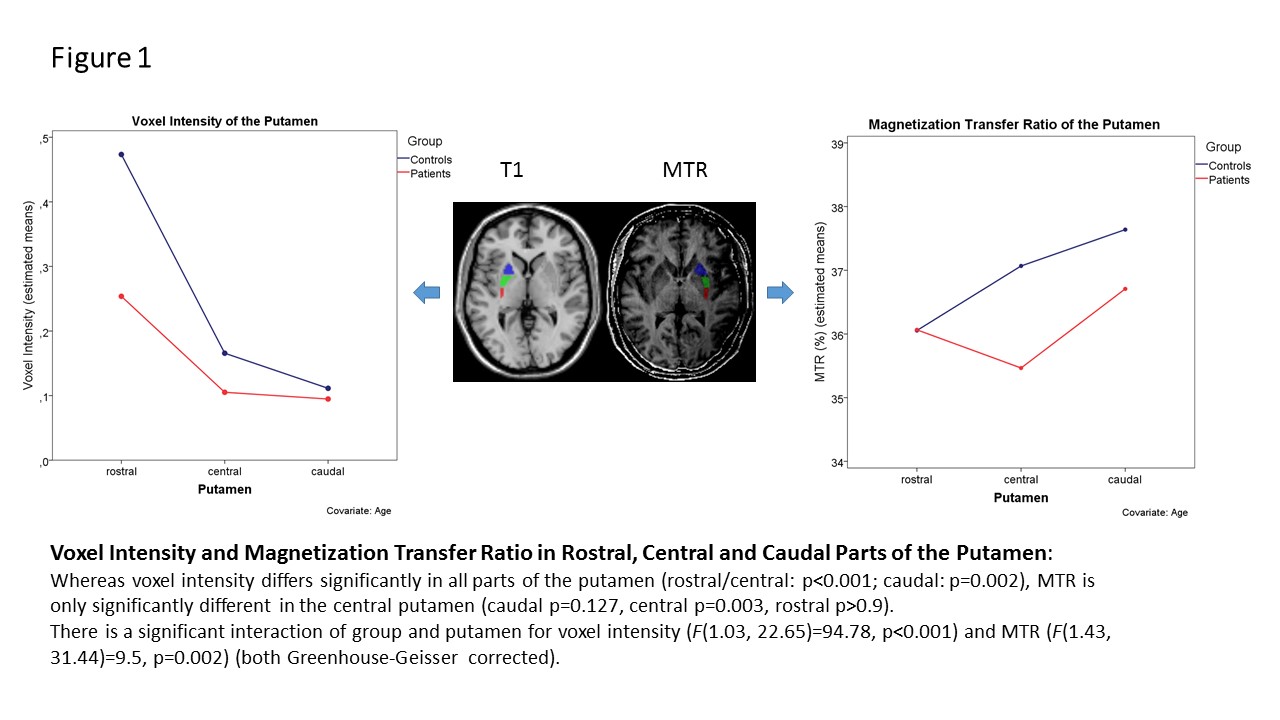
Results: In line with previous findings, putaminal voxel intensity as a marker of gray matter pathology was significantly lower in the patient group (p<0.001). Here, the reduction of voxel intensity followed a caudo-rostral gradient (caudal (-14.41%), central (-38.55%), and rostral putamen (-46.30%)). In contrast to these distinct abnormalities throughout the entire putamen, MTR values were reduced in the central putamen only and not in the other parts of the putamen. [figure1]
Conclusions: Putaminal atrophy in XDP is severe even after relatively short disease duration. T1 and MT imaging depict different aspects of neurodegeneration in XDP. In the rostral putamen, similar MT ratios in the presence of distinct atrophy upon T1 images may indicate a terminated neurodegenerative process whereas neurodegeneration is still ongoing in other parts of the putamen. Given the predominant motor phenotype of XDP, the observed caudo-rostral atrophy gradient is in contrast to the hypothesized preferential degeneration of the caudal sensorimotor putamen. Further multimodal imaging of the basal ganglia is warranted to better understand these findings and to interpret the significance of MT imaging in XDP.
To cite this abstract in AMA style:
H. Hanßen, M. Heldmann, C. Diesta, R. Rosales, A. Domingo, T. Münte, C. Klein, N. Brüggemann. Putaminal Atrophy Gradient in X-linked Dystonia-Parkinsonism [abstract]. Mov Disord. 2017; 32 (suppl 2). https://www.mdsabstracts.org/abstract/putaminal-atrophy-gradient-in-x-linked-dystonia-parkinsonism/. Accessed December 13, 2025.« Back to 2017 International Congress
MDS Abstracts - https://www.mdsabstracts.org/abstract/putaminal-atrophy-gradient-in-x-linked-dystonia-parkinsonism/

