Session Information
Date: Monday, October 8, 2018
Session Title: Parkinson's Disease: Pathophysiology
Session Time: 1:15pm-2:45pm
Location: Hall 3FG
Objective: To examine whether clinical, dopamine transporter (DAT) imaging and cerebrospinal fluid (CSF) markers can be used to predict the later development of FOG individually and in combination in patients with newly diagnosed Parkinson’s disease (PD).
Background: Various clinical factors have been suggested as predictors of FOG, but their predictive value remains unclear. Although presynaptic striatal dopamine depletion and CSF β-amyloid 1-42 (Aβ42) is considered to have a role in the development of FOG, they have not been investigated as a means of predicting FOG.
Methods: This retrospective cohort study included a total of 393 patients with newly diagnosed PD without FOG at baseline. Data were obtained from the Parkinson’s Progression Markers Initiative (PPMI) database. Data up to 4 years of follow-up were included. The development of FOG was defined to be present if the score was 1 or higher either for the Movement Disorder Society Unified Parkinson’s Disease Rating Scale (MDS-UPDRS) item 2.13 or item 3.11 at any point during the follow-up period. Cox regression models were performed to identify the factors predictive of FOG. Based on these results, we constructed a predictive model for the development of FOG.
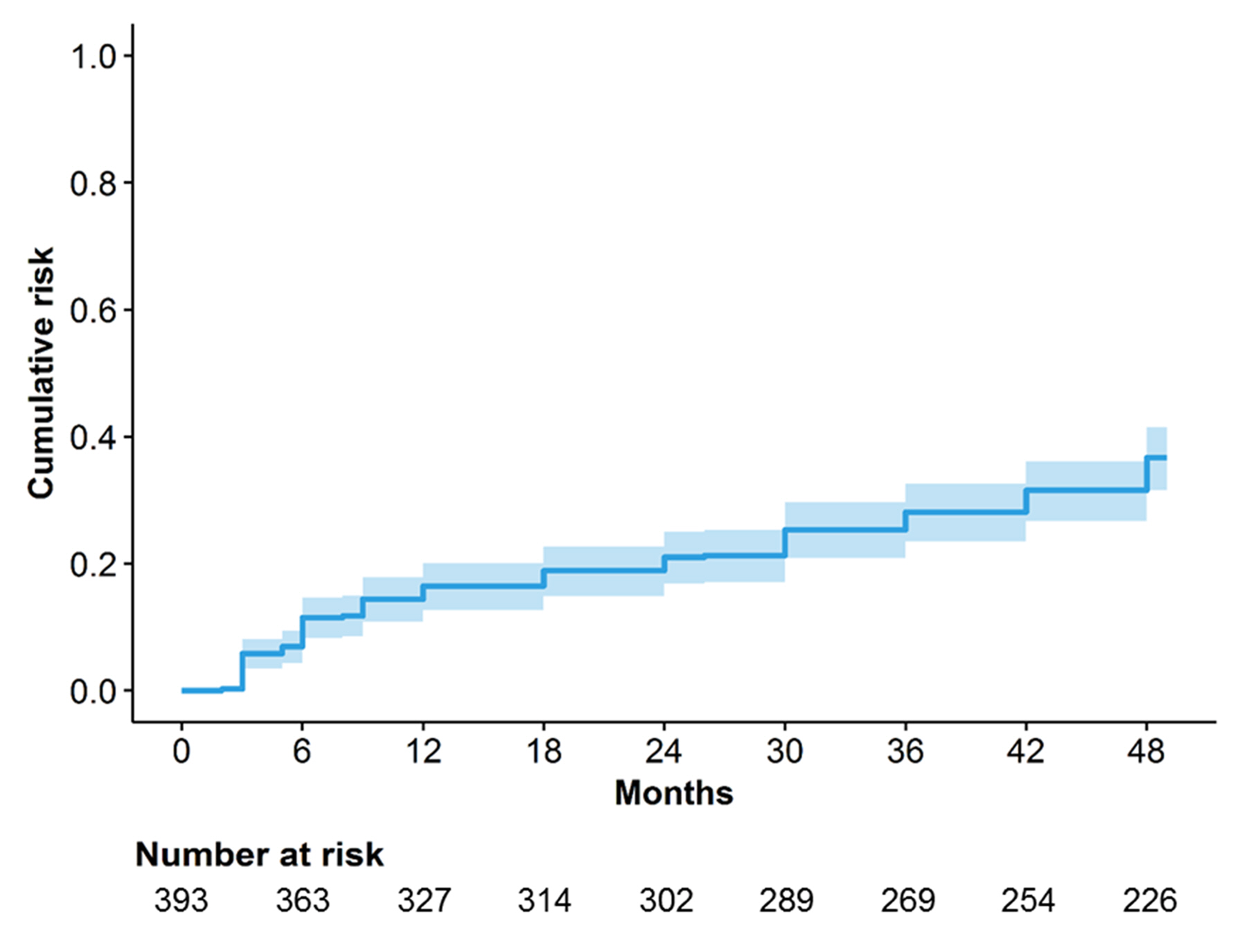
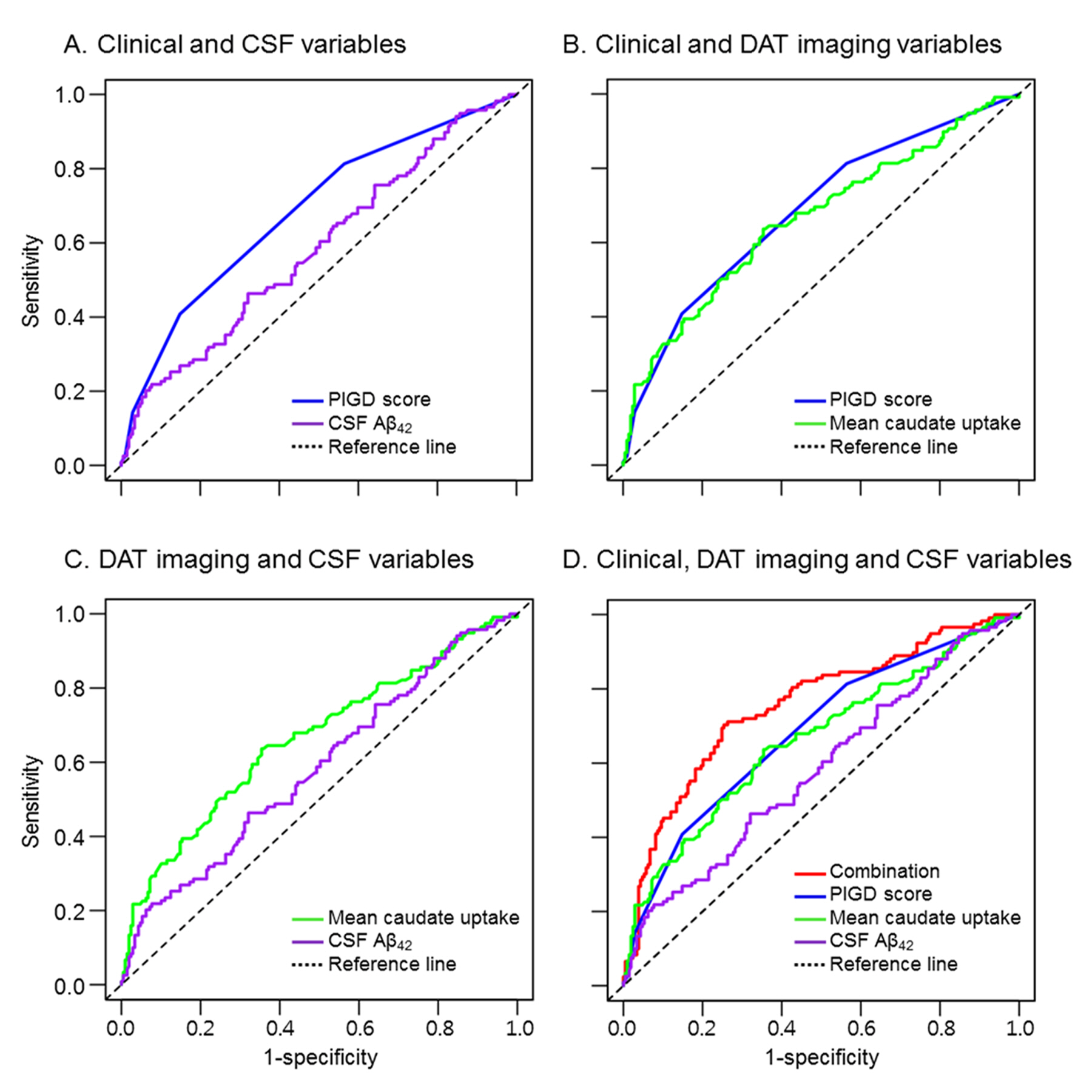
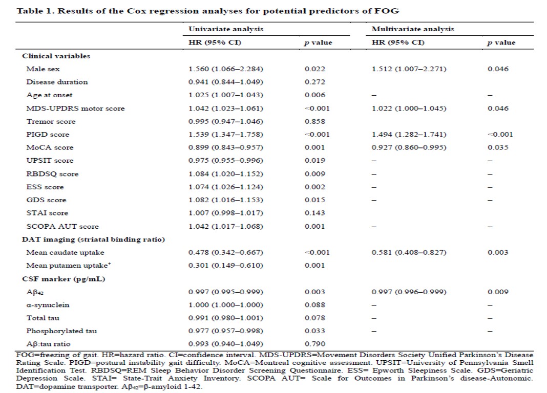
Results: During a median follow-up of 4.0 years (mean 3.0 years), 136 subjects developed FOG, and its cumulative incidence was 17, 21, 28, and 37% at 1-, 2-, 3- and 4-year follow-up, respectively [Figure 1]. The development of FOG was associated with postural instability gait difficulty (PIGD) score (hazard ratio [HR] 1.494; confidence interval [CI] 1.282–1.741; p<0.001), caudate DAT uptake (HR 0.581; 95% CI 0.408–0.827; p=0.003), CSF Aβ42 (HR 0.997; 95% CI 0.996–0.999; p=0.009) and, to a lesser extent, male sex (HR 1.512; 95% CI 1.007–2.271; p=0.046), MDS-UDPRS motor score (HR 1.022; 95% CI 1.000–1.045; p=0.046), and Montreal Cognitive Assessment score (HR 0.927; 95% CI 0.860–0.995; p=0.035) [Table 1]. The combined model integrating the PIGD score, caudate DAT uptake and CSF Aβ42 achieved a better prediction accuracy (area under the curve 0.755; 95% CI 0.700–0.810) than any factor alone [Figure 2].
Conclusions: We found clinical, DAT imaging and CSF markers as predictors of FOG in patients with newly diagnosed PD. The development of FOG within 4 years after diagnosis of PD can be predicted with acceptable accuracy using our risk model.
To cite this abstract in AMA style:
R. Kim, J. Lee, H.J. Kim, A. Kim, B. Jeon, U.J. Kang, S. Fahn. Predictors of freezing of gait in newly diagnosed Parkinson’s disease: Clinical, dopamine transporter imaging and CSF markers in the PPMI cohort [abstract]. Mov Disord. 2018; 33 (suppl 2). https://www.mdsabstracts.org/abstract/predictors-of-freezing-of-gait-in-newly-diagnosed-parkinsons-disease-clinical-dopamine-transporter-imaging-and-csf-markers-in-the-ppmi-cohort/. Accessed April 26, 2025.« Back to 2018 International Congress
MDS Abstracts - https://www.mdsabstracts.org/abstract/predictors-of-freezing-of-gait-in-newly-diagnosed-parkinsons-disease-clinical-dopamine-transporter-imaging-and-csf-markers-in-the-ppmi-cohort/


