Session Information
Date: Monday, October 8, 2018
Session Title: Parkinson's Disease: Neuroimaging And Neurophysiology
Session Time: 1:15pm-2:45pm
Location: Hall 3FG
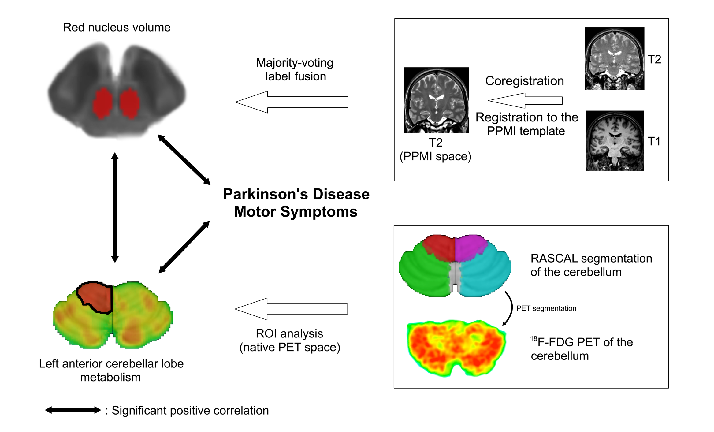
Objective: To investigate the involvement of the red nucleus (RN) and cerebellum in the motor symptoms of Parkinson’s disease (PD).
Background: Numerous studies have shown that the cerebellum is involved in PD, but its precise role remains unclear in this disease. As the red nucleus (RN) is intricately connected to the cerebellum, this structure has also been hypothesized to be involved in PD. Studies have reported greater RN volume in Parkinsonian patients compared with controls, and a positive correlation between disease severity and RN volume. However, these findings need to be verified, as sample sizes were small, resulting in poor statistical power.
Methods: We retrospectively examined 72 patients with PD. All patients had undergone whole-brain T1-weighted (T1w) and T2w MRI and 18F-fluorodeoxyglucose PET (18F-FDG PET). For each patient, RN volume was automatically estimated from the T2w MRI scan, and cerebellar metabolism was extracted from the 18F-FDG PET scan for each of the lobes. We assessed the correlations between RN volume, cerebellar metabolism and PD motor symptoms, as measured by the Unified Parkinson’s Disease Rating Scale Part III off medication (UPDRS-III OFF).
Results: We observed a significant positive correlation between the RN volume and the UPDRS-III OFF score (squared-r = 0.09, p = 0.009), and between left anterior cerebellar lobe metabolism (which is involved in sensory-motor processes and motor skills learning) and the UPDRS-III OFF score (squared-r = 0.10, p = 0.001). In addition, we found a significant positive correlation between the RN volume and left anterior cerebellar lobe metabolism (squared-r = 0.07, p = 0.036). Nevertheless, the multivariate analysis showed that both RN volume (squared-r = 0.11, p = 0.035) and left anterior cerebellar lobe metabolism (squared-r = 0.06, p = 0.003) had significant independent effects on the UPDRS-III OFF score.
Conclusions: These results indicate that cerebello-rubral pathways, but also RN and cerebellum independently, play a role in the motor symptoms of PD, which is likely to be compensatory. Thus, they demonstrate the need for therapies targeting these structures.
References: 1. Wu T, Hallett M. The cerebellum in Parkinson’s disease. Brain 2013;136:696-709. 2. Colpan ME, Slavin KV. Subthalamic and red nucleus volumes in patients with Parkinson’s disease: do they change with disease progression? Parkinsonism Relat Disord 2010;16:398-403.
To cite this abstract in AMA style:
A. Bonnet, R. Sanford, A. Riou, S. Drapier, F. Le Jeune, M. Vérin, D.L. Collins. Parkinson’s disease motor symptoms are linked to red nucleus volume and cerebellar metabolism [abstract]. Mov Disord. 2018; 33 (suppl 2). https://www.mdsabstracts.org/abstract/parkinsons-disease-motor-symptoms-are-linked-to-red-nucleus-volume-and-cerebellar-metabolism/. Accessed December 19, 2025.« Back to 2018 International Congress
MDS Abstracts - https://www.mdsabstracts.org/abstract/parkinsons-disease-motor-symptoms-are-linked-to-red-nucleus-volume-and-cerebellar-metabolism/

