Session Information
Date: Saturday, October 6, 2018
Session Title: Cognitive Disorders
Session Time: 1:45pm-3:15pm
Location: Hall 3FG
Objective: In this ex-vivo study, we aimed to assess atrophy in regions and subregions of the Papez Circuit, a network important for memory function, in patients with Dementia with Lewy Bodies (DLB). Results were compare to patients with Alzheimer’s disease (AD) with the objective of understanding structural changes subserving memory deficits specific to DLB.
Background: DLB is the second most common neurodegenerative dementia in the elderly. Although the neuropsychological phenotype of DLB has been distinguished from AD on the basis of more pronounced executive and visuospatial dysfunction, it is well known that DLB can often also present with significant memory disturbance akin to AD[1]. Previous in vivo studies have shown relative preservation of medial temporal lobe structures including the hippocampus in DLB relative to AD[2]. However, the hippocampus is only one component of a distributed network of structures subserving memory known as the Papez circuit. Projections within the Papez circuit involve the hippocampus, mammillary bodies, anterior thalamus and cingulate cortex. Significant atrophy[3] and hypometabolism[4] has been found across this network of regions in AD patients but to the best of our knowledge atrophy in the Papez circuit is yet to be investigated in DLB.
Methods: 18 cases of AD (9 males, 9 females), 18 cases of DLB (14 males, 4 females) and 20 control cases (10 males, 10 females) were chosen for post-mortem evaluation. The subregions of the hippocampus, mammillary bodies, anterior thalamus, and cingulate cortex were analysed using standard volumetric techniques[5].
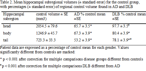
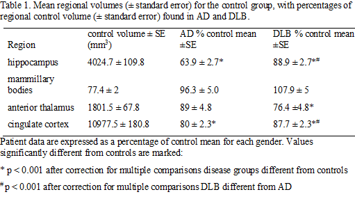
Results: There were significant differences in volumes of Papez memory circuit regions between groups (p<0.001). DLB cases had significantly more atrophy in the hippocampus, cingulate cortex and the anterior thalamus compared to controls. AD cases demonstrated significantly more volume loss than DLB and controls in all regions except the anterior thalamus which was similar to controls(Table 1). Subregion analysis revealed that hippocampal atrophy in DLB was concentrated in the body whilst AD patients had atrophy across all regions(Table 2). Cingulate cortex was differentially affected in DLB, with less posterior cingulate (PCC) atrophy compared to AD(Table 3).
Conclusions: Divergent patterns of atrophy within the memory circuits in DLB compared to AD and controls, with the anterior thalamus most affected in DLB and the hippocampus most affected in AD. We also found relative sparing of the PCC in DLB consistent with results from functional imaging[6]. Our findings suggest that the neural substrates of episodic memory impairment in DLB is subserved by a different pattern of memory network dysfunction to AD and provides clues towards potential in vivo imaging biomarkers that may differentiate between these conditions.
References: [1] Metzler-Baddeley, C. 2007. A review of cognitive impairments in dementia with Lewy bodies relative to Alzheimer’s disease and Parkinson’s disease with dementia. Cortex, 43, 583-600. [2] Burton, E.J., Barber, R., Mukaetova-Ladinska, E.B., Robson, J., Perry, R.H., Jaros, E., Kalaria, R.N., O-Brien, J.T. 2009. Medial temporal lobe atrophy on MRI differentiates Alzheimer’s disease from dementia with Lewy bodies and vascular cognitive impairment: a prospective study with pathological verification of diagnosis. Brain, 132, 195-203. [3] Callen, D.J., Black, S.E., Gao, F., Caldwell, C.B., Szalai, J.P. 2001. Beyond the hippocampus: MRI volumetry confirms widespread limbic atrophy in AD. Neurology, 57, 1669-74. [4] Nestor, P.J., Fryer, T.D., Smielewski, P., Hodges J.R. 2003. Limbic hypometabolism in Alzheimer’s disease and mild cognitive impairment. Ann Neurol, 54, 343-51. [5] Halliday, G.M., Double, K.L., Macdonald, V., Kril, J.J. 2003. Identifying severely atrophic cortical subregions in Alzheimer’s disease. Neurobiol Aging, 24, 797-806. [6] Iizuka, T., Kametama, M. 2016. Cingulate island sign on FDG-PET is associated with medial temporal lobe atrophy in dementia with Lewy bodies. Ann Nucl Med, 30(6): 421-9.
To cite this abstract in AMA style:
E. Matar, S. Wong, R. Tan, O. Piguet, M. Hornberger, J. Krill, J. Hodges, G. Halliday. Papez circuit atrophy in Dementia with Lewy Bodies [abstract]. Mov Disord. 2018; 33 (suppl 2). https://www.mdsabstracts.org/abstract/papez-circuit-atrophy-in-dementia-with-lewy-bodies/. Accessed April 26, 2025.« Back to 2018 International Congress
MDS Abstracts - https://www.mdsabstracts.org/abstract/papez-circuit-atrophy-in-dementia-with-lewy-bodies/


