Session Information
Date: Saturday, October 6, 2018
Session Title: Surgical Therapy: Parkinson's Disease
Session Time: 1:45pm-3:15pm
Location: Hall 3FG
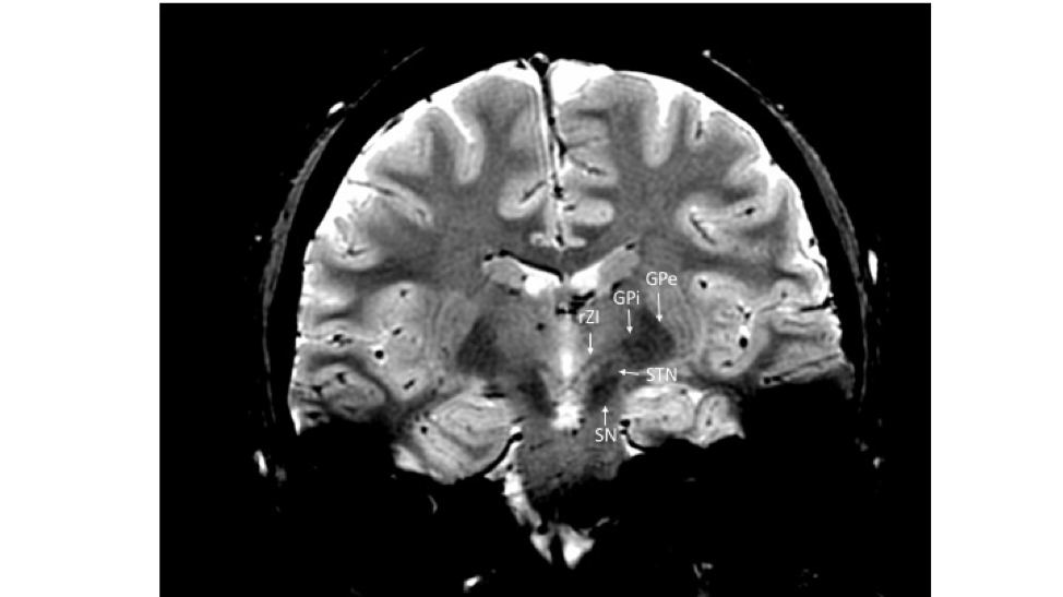
Objective: To improve upon imaging of the rostral zona incerta (rZI) allowing for direct targeting during deep brain stimulation.
Background: DBS of the subthalamic nucleus (STN) is an effective treatment for Parkinson’s disease (PD); however, unwanted, adverse effects may occur. Recently, the rZI has been proposed as a potential alternative DBS target for the treatment of PD. This structure is not reliably imaged with standard MRI sequences preventing direct stereotactic targeting. In this study, we present pilot data demonstrating the use of a novel 2D coronal gradient echo (2DGRE) sequence which allows consistent visualization of the rZI in both control and PD populations at 3.0 Tesla, not previously reported in the literature.
Methods: Six PD patients and four healthy control subjects had 2DGRE sequences performed to visualize the rZI. Comparisons in ability to visualize the rZI was made with T1 and T2 MRI sequences in the PD patients who underwent 2DGRE imaging as well as in an additional five PD patients. Imaging of all 15 subjects was independently and blind-reviewed by a neuroradiologist and a movement disorder neurologist, utilizing a 3 point Likert scale rating bilateral rZI visualization (0-nonvisualized, 1-partially visualized, 2-clearly visualized).
Results: There was high inter rater agreement between reviewers in scoring visualization of the rZI using the 2DGRE sequences in PD patients of 83% and in healthy subjects of 100%. The rZI was visualized in all individuals who underwent 2DGRE imaging [figure 1]. The rZI was better evident in healthy subjects compared with PD patients with a mean Likert score of 2 compared to 1.4, respectively. The rZI was not able to be visualized with conventional T1 and T2 MRI sequences.
Conclusions: We demonstrate promising initial data suggesting that a 2DGRE sequence can image the rZI at 3.0 Tesla in PD patients. Prior short reports have successfully imaged this small structure in healthy controls. Improved visualization will allow direct targeting of the rZI for electrode placement during DBS.
References: Merello M, et al., Mov Disord. 2006 Jul;21(7):937-43. Kerl HU, et al. Clin Neuroradiol. 2012 Mar;22(1):55-68.
To cite this abstract in AMA style:
A. Thaker, K. Reddy, J. Thompson, P. David-Gerecht, A. Abosch, D. Kern. Optimized Imaging at 3.0 T of the Rostral Zona Incerta (rZI) for Deep Brain Stimulation (DBS) in Parkinson’s Disease (PD) [abstract]. Mov Disord. 2018; 33 (suppl 2). https://www.mdsabstracts.org/abstract/optimized-imaging-at-3-0-t-of-the-rostral-zona-incerta-rzi-for-deep-brain-stimulation-dbs-in-parkinsons-disease-pd/. Accessed January 5, 2026.« Back to 2018 International Congress
MDS Abstracts - https://www.mdsabstracts.org/abstract/optimized-imaging-at-3-0-t-of-the-rostral-zona-incerta-rzi-for-deep-brain-stimulation-dbs-in-parkinsons-disease-pd/

