Session Information
Date: Wednesday, September 25, 2019
Session Title: Neuroimaging
Session Time: 1:15pm-2:45pm
Location: Les Muses Terrace, Level 3
Objective: To explore the pattern of activation within the motor network during the movement and resting phases of the tapping task in Parkinson’s disease (PD) before and after the levodopa challenge.
Background: Research investigating brain activity changes with levodopa treatment in PD using a functional MRI yielded variable activation patterns at the subcortical level [1-4] These conflicting results suggest a differential role of basal ganglia depending on the experimental context.
Method: The study included 32 patients with sporadic PD in an advanced stage with motor fluctuations (6 fem, HY II-III, age 56.1±7.7y, disease duration 12.2±2.5y, levodopa treatment duration 9.0±3.0y, mean±SD). A block-based motor paradigm was used with consecutive finger tapping movement and rest epochs, each lasting 10s. During tapping epochs (TAP), 10 pacing movement cues were presented with a frequency of 1 Hz. A functional MRI was performed on a 1.5T scanner (Siemens, Erlangen, Germany) with gradient-echo echo-planar imaging sequence (RT=1s; ET=54ms). Ten oblique slices were acquired along the central sulcus, covering the primary sensorimotor cortex and the basal ganglia. A two-by-two factorial design with within-subject factors ‘Hand’ (RIGHT/LEFT) and ‘Levodopa’ (OFF/ON) resulted in 4 scanning sessions for each patient. Significant results were reported with a family-wise error (FWE) correction at the voxel-level with p<0.05.
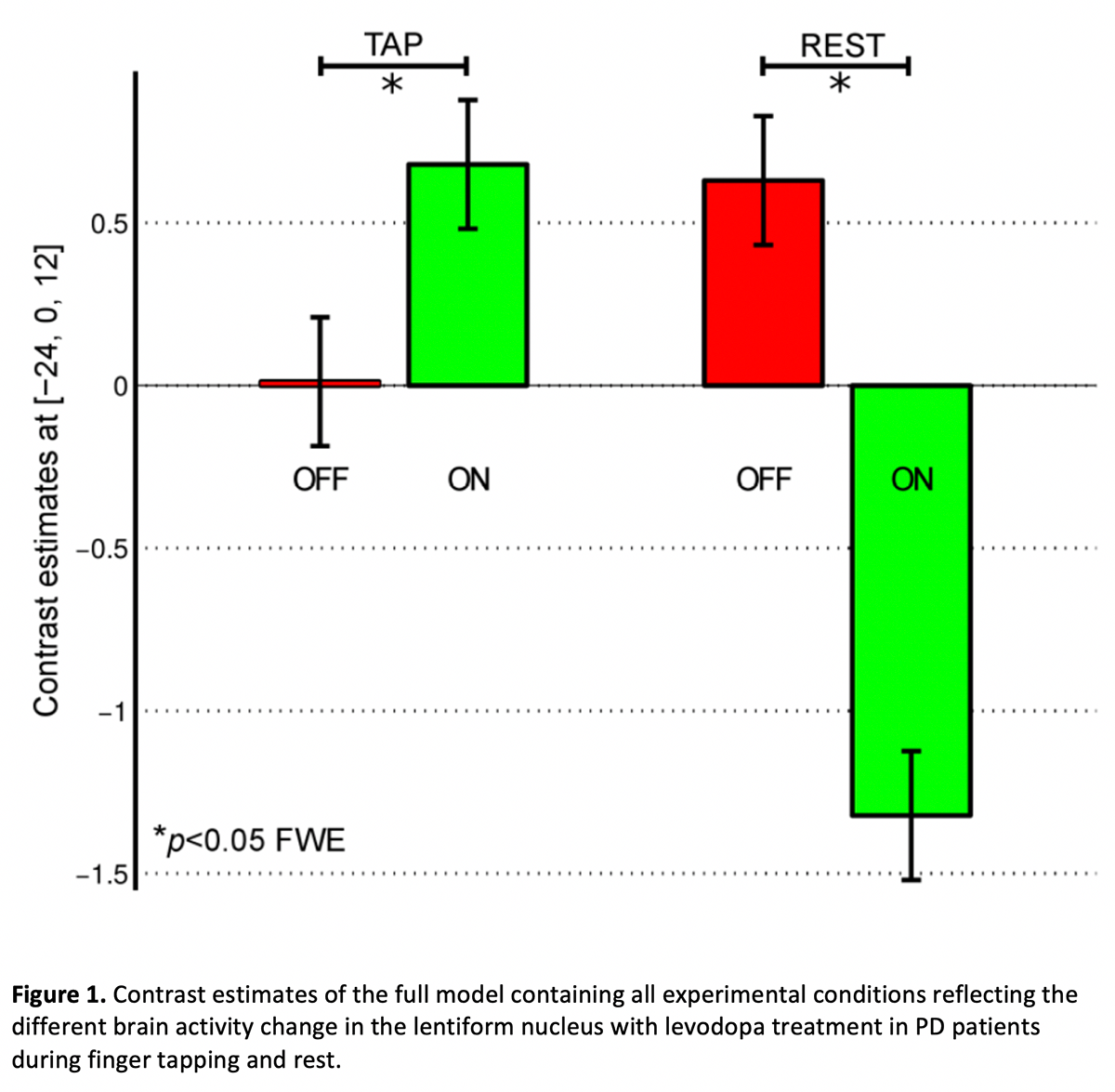
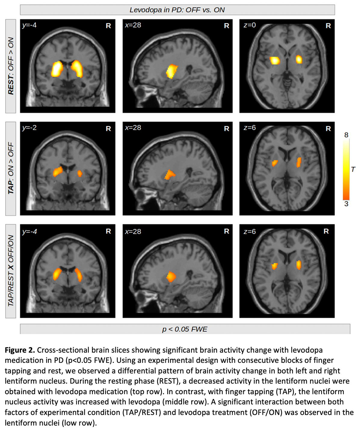
Results: There was a differential pattern of lentiform nucleus activity change with levodopa medication in the TAP and the REST phases (Fig 1). While in the TAP phase, we observed a significant increase of activity (ON>OFF, Fig 2, top row), in the REST phase, we observed a decrease of activity with levodopa administration (OFF>ON, Fig 2, middle row). [figure1][figure2]
Conclusion: We obtained an opposing pattern of brain activity with dopaminergic medication in PD patients depending on the phase of the movement task. The lentiform nuclei at rest were underactive in the ON compared to OFF state but their activity increased with the levodopa challenge when externally cued finger tapping was performed. This suggests a fundamental difference in the involvement of resting and active motor networks in PD depending on the actual dopamine level in the basal ganglia.Supported by the grant GACR 16-13323S.
References: 1. Kraft et al. (2009). Levodopa-induced striatal activation in Parkinson’s disease: a functional MRI study. Parkinsonism Relat Disord. 2009 Sep;15(8):558-63. 2. Kwak et al. (2012). l-DOPA changes ventral striatum recruitment during motor sequence learning in Parkinson’s disease. Behav Brain Res. 2012 Apr 21;230(1):116-24. 3. Maillet et al. (2012). Levodopa effects on hand and speech movements in patients with Parkinson’s disease: a FMRI study. PLoS One. 2012;7(10):e46541. 4. Holiga et al. (2014). Motor matters: tackling heterogeneity of Parkinson’s disease in functional MRI studies. PLoS One. 2013;8(2):e56133.
To cite this abstract in AMA style:
R. Jech, S. Holiga, F. Růžička, H. Möller, J. Roth, M. Schroeter, E. Růžička, K. Mueller. Opposing effects of levodopa on basal ganglia activity in Parkinson’s disease during motion and rest [abstract]. Mov Disord. 2019; 34 (suppl 2). https://www.mdsabstracts.org/abstract/opposing-effects-of-levodopa-on-basal-ganglia-activity-in-parkinsons-disease-during-motion-and-rest/. Accessed January 6, 2026.« Back to 2019 International Congress
MDS Abstracts - https://www.mdsabstracts.org/abstract/opposing-effects-of-levodopa-on-basal-ganglia-activity-in-parkinsons-disease-during-motion-and-rest/


