Category: Parkinson's Disease: Neuroimaging
Objective: The goal of this work is to image the microvasculature of the midbrain, in particular the substantia nigra in Parkinson’s disease (PD) patients using an ultra-small super-paramagnetic iron oxide (USPIO) contrast agent, ferumoxytol.
Background: Animal and human studies have shown that micro-vascular dysfunction is implicated in a variety of neurodegenerative diseases including PD. [1-4] Micro-vascular abnormalities may precede or accompany midbrain dopaminergic neuronal loss in PD patients. [3,4] A limited number of autopsy studies have indicated the presence of abnormal neuro-vasculature in the substantia nigra pars compacta. [4,5]
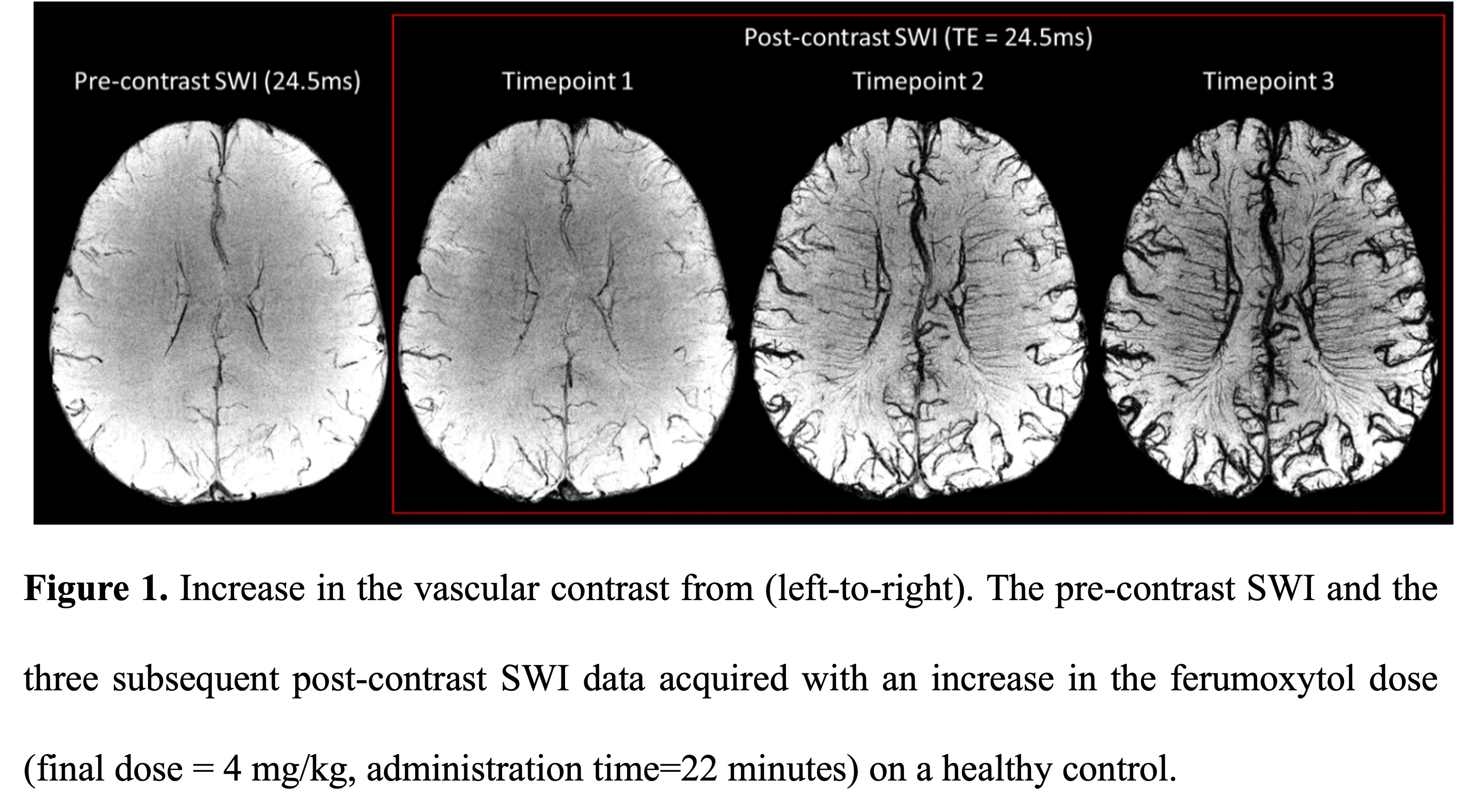
Method: This is a prospective study with ongoing enrolment of subjects with PD and age and sex matched healthy controls (HC). Both groups are being scanned at 3T utilizing MICRO (Microvascular In-vivo Contrast Revealed Origins) imaging. A low dose of 4mg/kg of ferumoxytol, an USPIO agent is used as contrast. The high susceptibility of this agent leads to large dephasing effects aiding in detection of vascular abnormalities at the micro-level (50-100 µm). [6,7] This 3D susceptibility weighted imaging (SWI) protocol has a resolution of 0.22×0.44×1mm3 and is acquired at four timepoints: once pre-contrast and three post-contrast acquisitions at 11-minute intervals during a gradual injection of ferumoxytol. The pre-contrast data highlights both major arteries and veins separately, whereas the post-contrast data highlights all vessels. [7] A 3D vessel density map is generated for direct visual inspection and quantitative measurements of both micro-arterial and micro-venous systems in the midbrain is done.
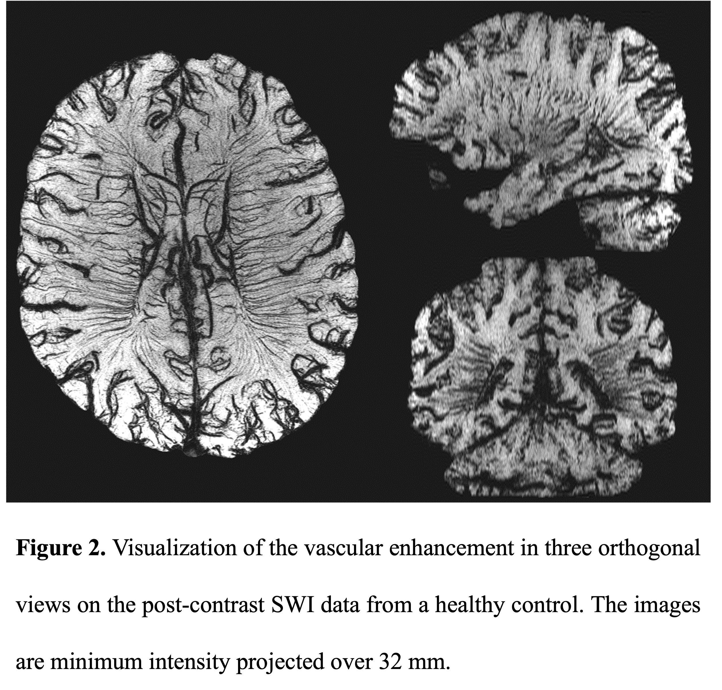
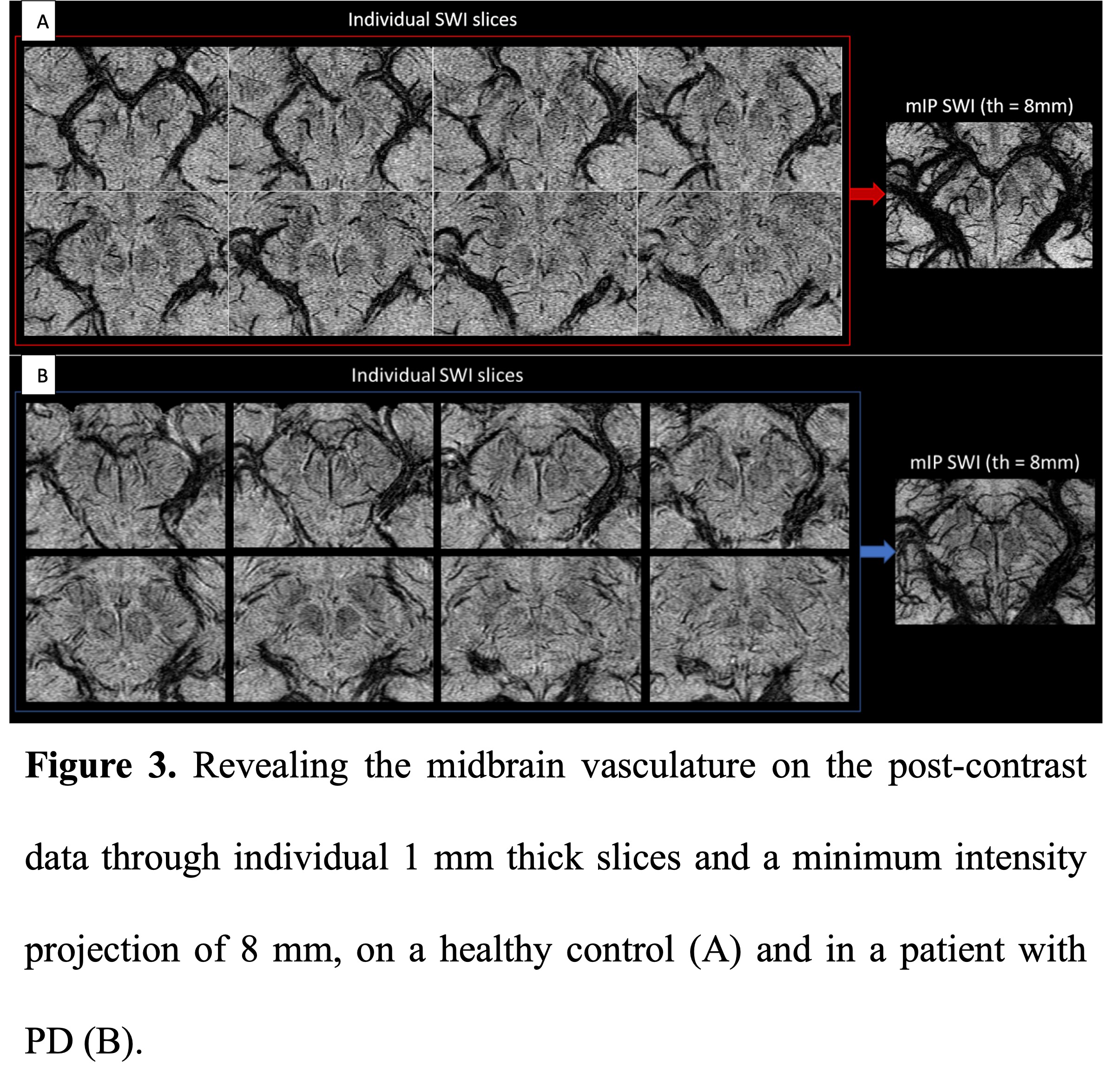
Results: Figures 1 and 2 show the vascular enhancement due to ferumoxytol as a function of time during the injection and then in different orientations for the last time point in a HC. Detailed midbrain micro-vasculature mapping using ferumoxytol guided increased susceptibility in a HC and PD patient is also shown (Figure 3A and B).
Conclusion: A detailed analysis of the micro-vascular architecture and vascular density is possible with MICRO imaging. The microcirculation plays a critical role in neuronal health. Therefore, any alteration in the micro-vascular environment of dopaminergic neurons in the Substantia nigra in PD patients could impact the metabolic equilibrium thereby triggering neurodegeneration. In the future, comparison of quantitative imaging biomarkers between PD patients and healthy controls can be studied.
References: 1. Kelleher RJ, Soiza RL. Evidence of endothelial dysfunction in the development of Alzheimer’s disease: is Alzheimer’s a vascular disorder? American journal of cardiovascular disease. 2013;3(4):197.
2. Zlokovic BV. The blood-brain barrier in health and chronic neurodegenerative disorders. Neuron. 2008;57(2):178-201.
3. Bradaric BD, Patel A, Schneider JA, Carvey PM, Hendey B. Evidence for angiogenesis in Parkinson’s disease, incidental Lewy body disease, and progressive supranuclear palsy. Journal of neural transmission. 2012;119(1):59-71.
4. Faucheux BA, Agid Y, Hirsch EC, Bonnet AM. Blood vessels change in the mesencephalon of patients with Parkinson’s disease. The Lancet. 1999;353(9157):981-2.
5. Issidorides MR. Neuronal vascular relationships in the zona compacta of normal and parkinsonian substantia nigra. Brain Research. 1971;25(2):289-99.
6. Shen YM, Zheng WL, Cheng YC, et al. USPIO high resolution neurovascular imaging in a rat stroke model of transient middle cerebral artery occlusion. Chinese Journal of Magnetic Resonance. 2014;31(1):20-31.
7. Buch S, Wang Y, Park MG, et al. Subvoxel vascular imaging of the midbrain using USPIO-Enhanced MRI. Neuroimage. 2020;220:117106.
To cite this abstract in AMA style:
S. Sharma, S. Buch, D. Reese, M. Haacke, M. Jog. Nigral microvasculature imaging in Parkinson’s disease [abstract]. Mov Disord. 2022; 37 (suppl 2). https://www.mdsabstracts.org/abstract/nigral-microvasculature-imaging-in-parkinsons-disease/. Accessed April 26, 2025.« Back to 2022 International Congress
MDS Abstracts - https://www.mdsabstracts.org/abstract/nigral-microvasculature-imaging-in-parkinsons-disease/


