Category: Parkinson's Disease: Neuroimaging
Objective: To compare the signal intensity of the substantia nigra and locus coeruleus on Magnetization Transfer 7T MRI between PD patients and healthy controls (HC) and to explore its association with cognitive performance in PD patients.
Background: Neuromelanin related signal changes in catecholaminergic nuclei are considered one of the most promising MRI biomarkers in Parkinson’s disease [1]. Until now, the vast majority of studies have focussed on the substantia nigra, while evidence suggests that lower neuromelanin related signal in early-stage Parkinson’s disease (PD) might be more prominent in the locus coeruleus [2]. Ultra-high field MRI techniques improve the visualisation of these small brainstem regions and might be suitable to detect neuromelanin related signal changes as a PD biomarker [3].
Method: This study was conducted using data from the TRACK-PD study, a longitudinal 7T-MRI study in PD patients. 78 early-stage PD patients and 36 HC were included. A mask for the substantia nigra and locus coeruleus was automatically segmented and manually corrected. Neuromelanin related signal intensity of the substantia nigra and locus coeruleus was compared between PD and HC.
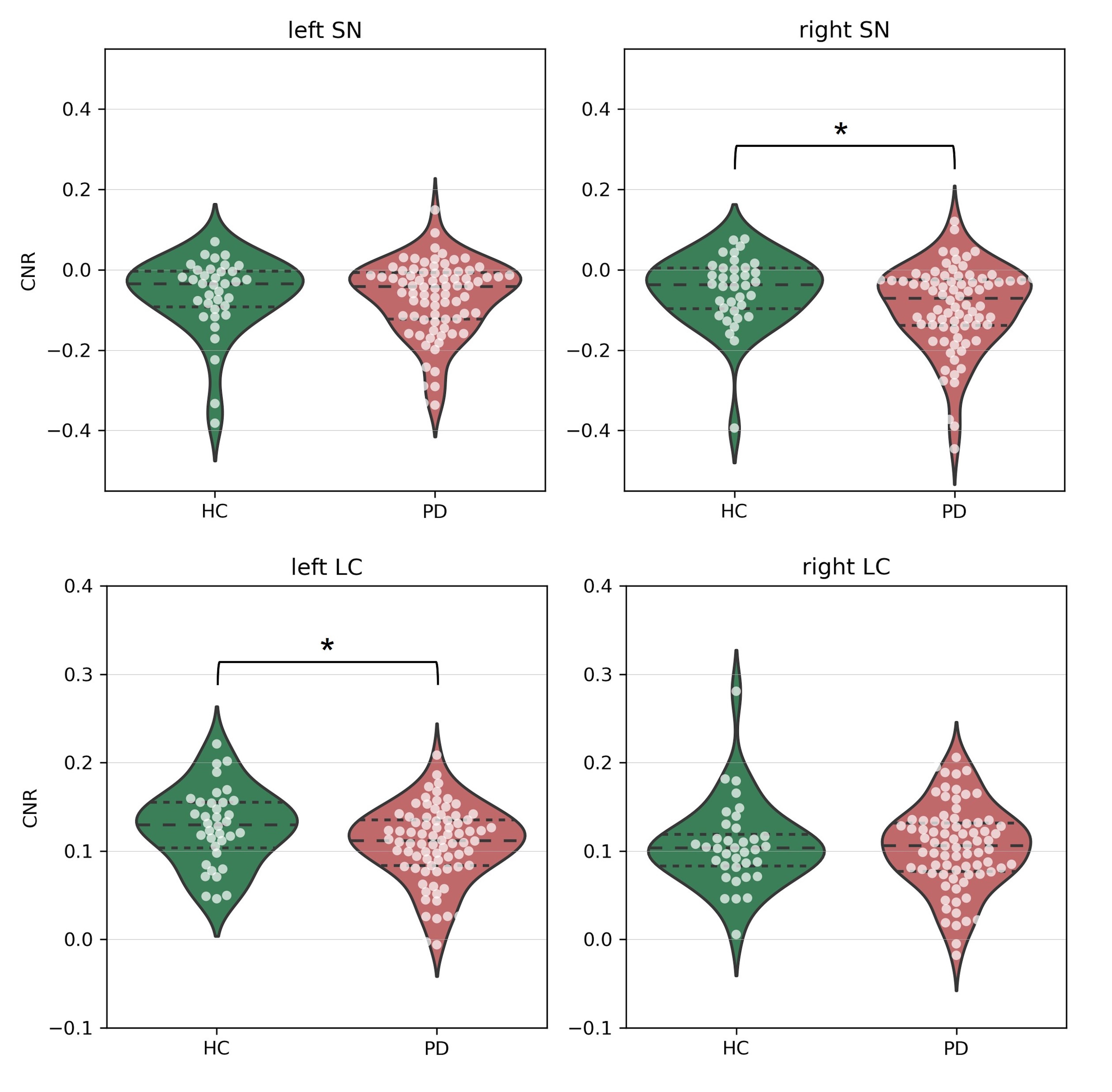
Results: PD participants showed a significantly lower contrast-to-noise ratio in the right substantia nigra (p = 0.029) and left locus coeruleus (p = 0.027) (Figure 1). After adding age as a confounder, the contrast-to-noise ratio of the right substantia nigra did not significantly differ anymore between PD and HC (p = 0.055). In addition, a significant positive correlation was found between the substantia nigra contrast-to-noise ratio and memory function.
Conclusion: Our findings confirm that neuromelanin related signal intensity of the locus coeruleus differs between early-stage PD patients and HC. No significant difference was found in the substantia nigra. This supports the theory of bottom-up disease progression in PD. Furthermore, loss of substantia nigra integrity might contribute to working memory deficits in PD. Future studies should focus on longitudinal signal changes in the locus coeruleus and substantia nigra in order to further elucidate the pathophysiological mechanisms of PD.
References: [1] Lehericy S, Vaillancourt DE, Seppi K, Monchi O, Rektorova I, Antonini A, et al. The role of high-field magnetic resonance imaging in parkinsonian disorders: Pushing the boundaries forward. Mov Disord. 2017;32(4):510-25.
[2] Isaias IU, Trujillo P, Summers P, Marotta G, Mainardi L, Pezzoli G, et al. Neuromelanin Imaging and Dopaminergic Loss in Parkinson’s Disease. Front Aging Neurosci. 2016;8:196.
[3] Prange S, Metereau E, Thobois S. Structural Imaging in Parkinson’s Disease: New Developments. Curr Neurol Neurosci Rep. 2019;19(8):50.
To cite this abstract in AMA style:
A. Wolters, M. Heijmans, N. Priovoulos, H. Jacobs, A. Postma, Y. Temel, M. Kuijf, S. Michielse. Neuromelanin related 7T MRI of the locus coeruleus differs between Parkinson’s disease and controls [abstract]. Mov Disord. 2023; 38 (suppl 1). https://www.mdsabstracts.org/abstract/neuromelanin-related-7t-mri-of-the-locus-coeruleus-differs-between-parkinsons-disease-and-controls/. Accessed December 31, 2025.« Back to 2023 International Congress
MDS Abstracts - https://www.mdsabstracts.org/abstract/neuromelanin-related-7t-mri-of-the-locus-coeruleus-differs-between-parkinsons-disease-and-controls/

