Category: Rare Genetic and Metabolic Diseases
Objective: To determine the anatomical basis for cognitive deficits in WD.
Background: Cognitive impairment is common in neurological presentations of Wilson’s disease (WD).1 Various cognitive domains can be affected and subclinical deficits have been reported in hepatic presentations.2 Associations with imaging abnormalities have not been systematically tested.
Method: We compared scores for 16 neuropsychological tests between patients with neurological (n=23) and hepatic (n=17) presentations and with normative data. Associations with Unified WD Rating Scale (UWDRS) neurological examination subscores were examined. Quantitative, whole-brain, multi-modal imaging analyses were used to identify associations with neuroimaging abnormalities in chronically-treated, stable patients (n=35), as previously described.3Analyses were performed with and without including UWDRS neurological examination subscores as a covariate.
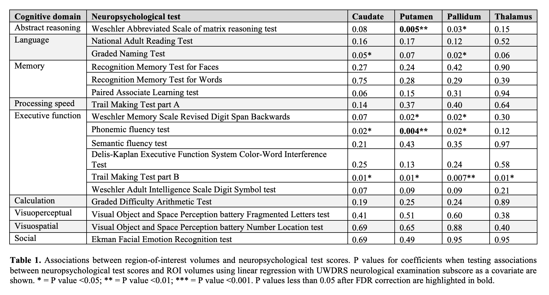
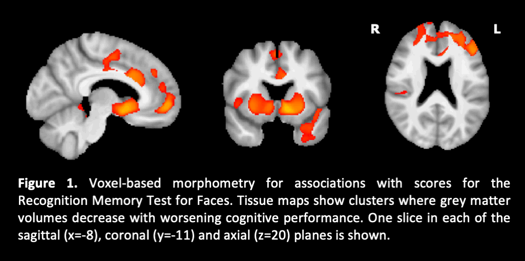
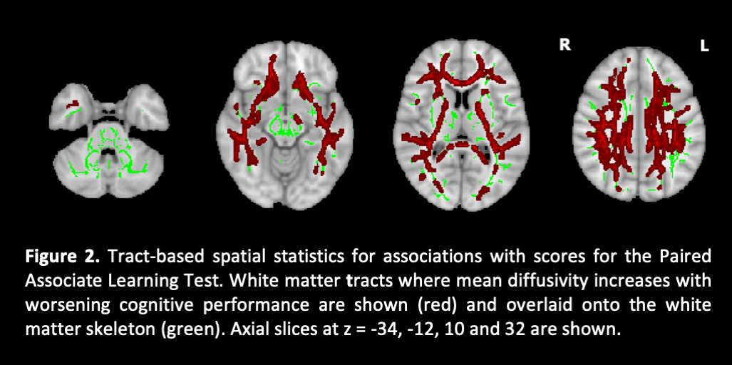
Results: Abstract reasoning, executive function, processing speed and visuospatial function scores were lower in patients with neurological presentations and correlated with UWDRS neurological examination subscores (P<0.05). Deficits in abstract reasoning and phonemic fluency were associated with lower putamen volumes even after controlling for neurological severity (region-of-interest volumetric analysis, FDR-correctedP<0.05; table 1). Around half of patients with hepatic presentations had deficits in memory for faces, cognitive flexibility or associative learning. These were associated with widespread cortical atrophy (voxel-based morphometry, FWE-correctedP<0.05; figure 1) and/or white matter diffusion abnormalities (tract-based spatial statistics, FWE-correctedP<0.05; figure 2).
Conclusion: Subtle cognitive deficits that appear to be common in patients with hepatic presentations represent a distinct neurological phenotype associated with diffuse cortical and white matter pathology. This may precede the classical phenotype characterised by movement disorders and executive dysfunction associated with basal ganglia damage. A binary disease classification may no longer be appropriate.
References: 1. Seniów J, Bak T, Gajda J, et al. Cognitive functioning in neurologically symptomatic and asymptomatic forms of Wilson’s disease. Mov Disord 2002;17(5):1077–83.
2. Tarter RE, Switala J, Carra J, et al. Neuropsychological impairment associated with hepatolenticular degeneration (Wilson’s disease) in the absence of overt encephalopathy. Int J Neurosci 1987;37(1-2):67–71.
3. Shribman S, Bocchetta M, Sudre CH, et al. Neuroimaging correlates of brain injury in Wilson’s disease: a multimodal, whole-brain MRI study. Brain 2021. https://doi.org/10.1093/brain/awab274.
To cite this abstract in AMA style:
S. Shribman, M. Burrows, R. Convery, M. Bocchetta, C. Sudre, J. Acosta-Cabronero, D. Thomas, G. Gillett, E. Tsochatzis, O. Bandmann, J. Rohrer, T. Warner. Neuroimaging correlates of cognitive deficits in Wilson’s disease [abstract]. Mov Disord. 2022; 37 (suppl 2). https://www.mdsabstracts.org/abstract/neuroimaging-correlates-of-cognitive-deficits-in-wilsons-disease/. Accessed December 30, 2025.« Back to 2022 International Congress
MDS Abstracts - https://www.mdsabstracts.org/abstract/neuroimaging-correlates-of-cognitive-deficits-in-wilsons-disease/


