Session Information
Date: Wednesday, September 25, 2019
Session Title: Neuroimaging
Session Time: 1:15pm-2:45pm
Location: Les Muses Terrace, Level 3
Objective: Aim of the study is to evaluate the presence of grey matter (GM) and electrocortical changes in Pd patients with MCI.
Background: Mild cognitive impairment (MCI) is a common non-motor features in Parkinson’s disease (PD), but the underlying pathological mechanism has not been fully understood. Neuroimaging and neurophysiological studies, showed the presence of atrophy and abnormal electrocortical activity in PD with cognitive decline, respectively. Voxel-based morphometry (VBM) and quantitative EEG analysis could be used to identify in vivo markers linked to the development of MCI in PD patients.
Method: From the PaCoS (Parkinson’s disease Cognitive impairment Study) cohort, a sample of PD patients with and without MCI were recruited, including those patients with one EEG recording and a T1-3D MRI, acquired at the same time. VBM analysis was performed and EEG signal epochs were analysed using Independent Component Analysis LORETA.
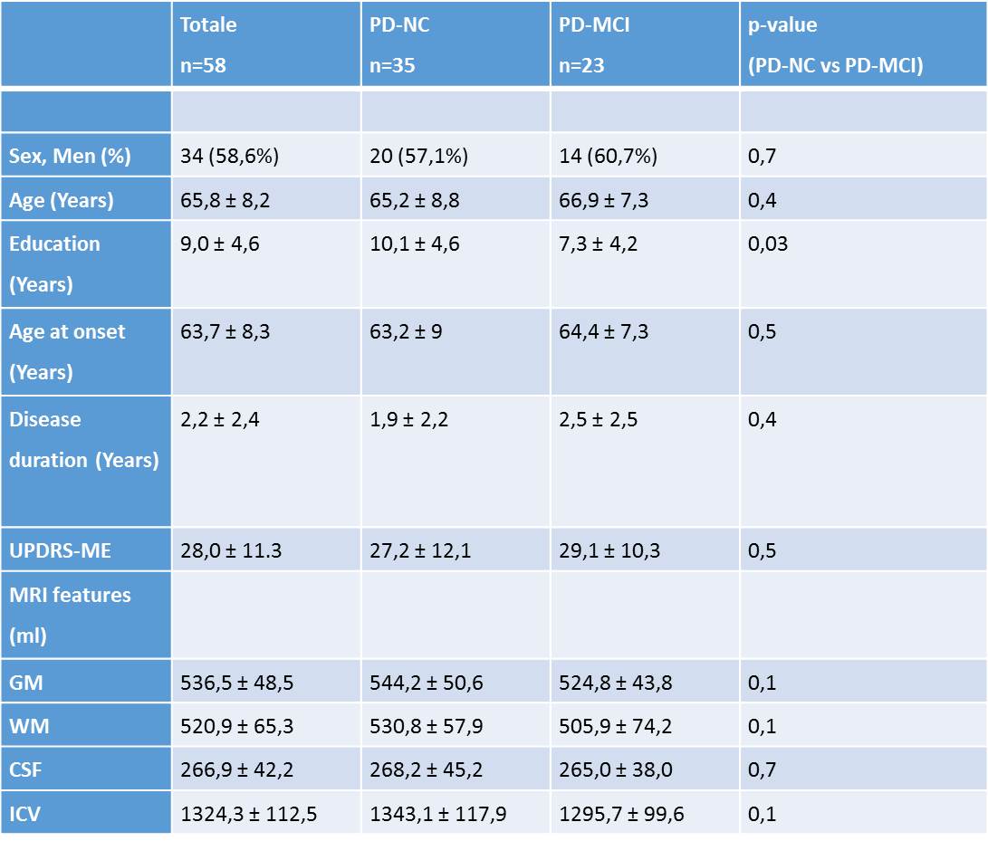
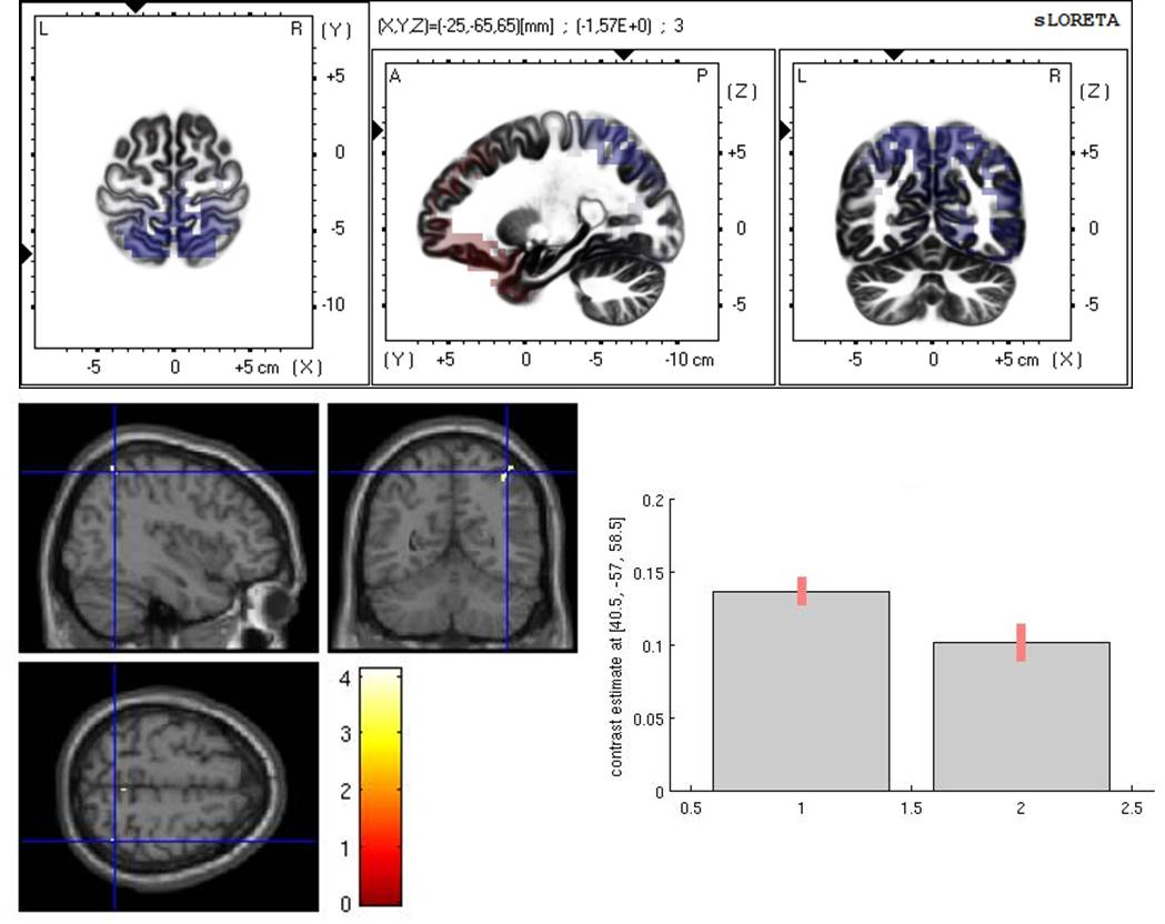
Results: Fifty-eight PD patients were enrolled, including 35 patients with normal cognition (PD-NC) and 23 PD with MCI (table1). PD-MCI showed reduction in GM density in a para-hippocampal areas, left temporal lobe, left cerebellum, precuneus, cingulate gyrus and right inferior parietal lobule. LORETA analysis revealed an increased network involving theta activity over the occipital lobe associated with a reduction over the frontal lobe. The parietal lobe showed a decreased network involving beta, delta and theta activity. Finally, a reduction of networks involving alfa and beta activity in the parietal lobule and inferior temporal gyrus was also found (figure1).
Conclusion: Our study showed, with a multimodal approach, the presence of widespread anatomical and electrocortical abnormalities with a common pattern in PD with MCI, involving mainly the parietal and temporal lobe. These results could be used as potential biomarkers of cognitive impairment in PD.
To cite this abstract in AMA style:
G. Donzuso, G. Giuliano, R. Monastero, R. Baschi, G. Mostile, A. Luca, C. Cicero, M. Zappia, A. Nicoletti. Neuroanatomical and Electrocortical Networks correlates in Parkinson’s Disease Patients with Mild Cognitive Impairment:The PaCoS Study [abstract]. Mov Disord. 2019; 34 (suppl 2). https://www.mdsabstracts.org/abstract/neuroanatomical-and-electrocortical-networks-correlates-in-parkinsons-disease-patients-with-mild-cognitive-impairmentthe-pacos-study/. Accessed April 21, 2025.« Back to 2019 International Congress
MDS Abstracts - https://www.mdsabstracts.org/abstract/neuroanatomical-and-electrocortical-networks-correlates-in-parkinsons-disease-patients-with-mild-cognitive-impairmentthe-pacos-study/


