Category: Parkinson's Disease: Neuroimaging
Objective: The main aims of this study were to characterize brain microstructure alterations in a cross-sectional cohort of Parkinson’s disease (PD) with DTI and assess associations of DTI outcomes with DAT availability.
Background: 39 PD subjects without dementia [19F, age (mean±sd): 64±7y, MDS-UPDRS Part III:27±15, Hoehn-Yahr:2±0] and 22 healthy controls (HC) [14F, age:45±15y] underwent diffusion weighted imaging (b=2000 s/mm2, 64 directions, TR=4100ms, TE=88ms) on a 3T MR scanner. A sub-cohort of 24 subjects (15 PD, 9 HC) underwent dynamic DAT PET imaging on the HRRT with [18F]FE-PE2I.
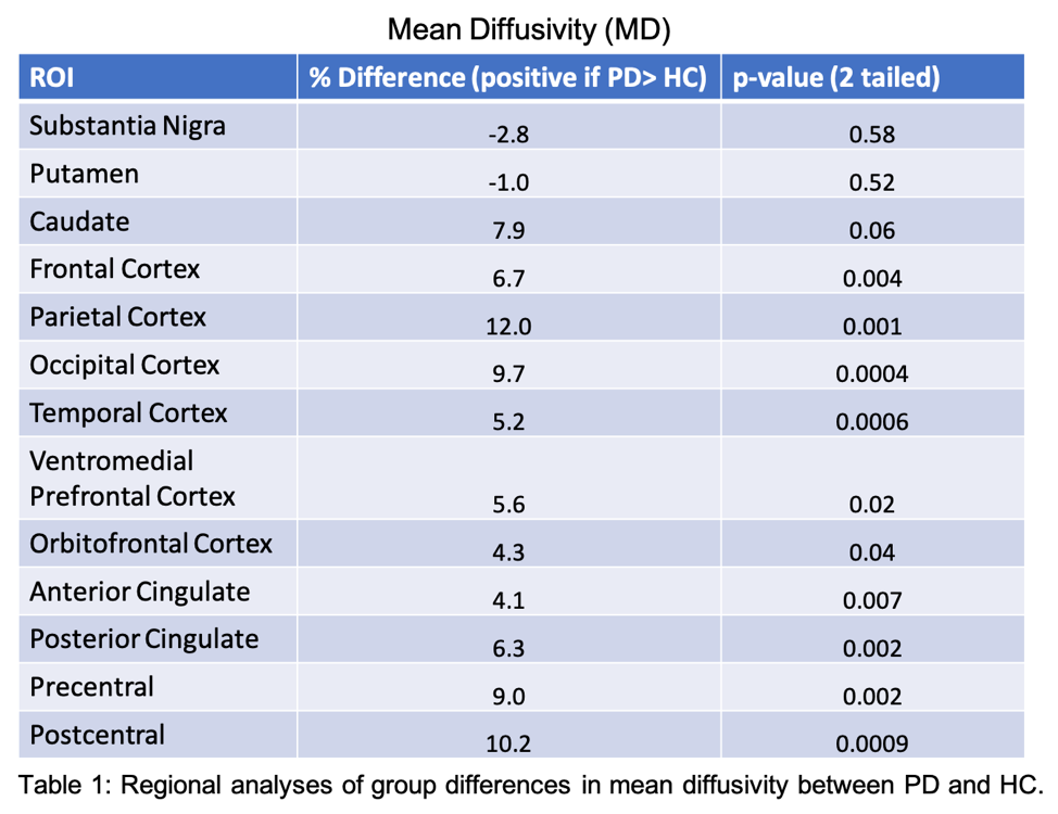
Method: Diffusion images, corrected for susceptibility and eddy current distortions, were fit to a tensor model (FDT, FSLv6.0.1). Mean diffusivity (MD) and fractional anisotropy (FA) were assessed in white matter (WM) tracts through tract-based spatial statistics (TBSS), and in gray matter, by defining regions of interest on a standard template for the caudate, putamen and several cortical areas (Table 1). For the substantia nigra (SN), an in-house template based on previous PET studies was used. Kinetic modeling of dynamic PET data (SRTM, ref=cerebellum) was used to estimate DAT binding potential (BPND) in the SN, caudate and putamen. In addition to group differences, regional correlations with MDS-UPDRS Part III and associations between DAT BPND and DTI measures in the SN, caudate and putamen were evaluated in PD subjects.
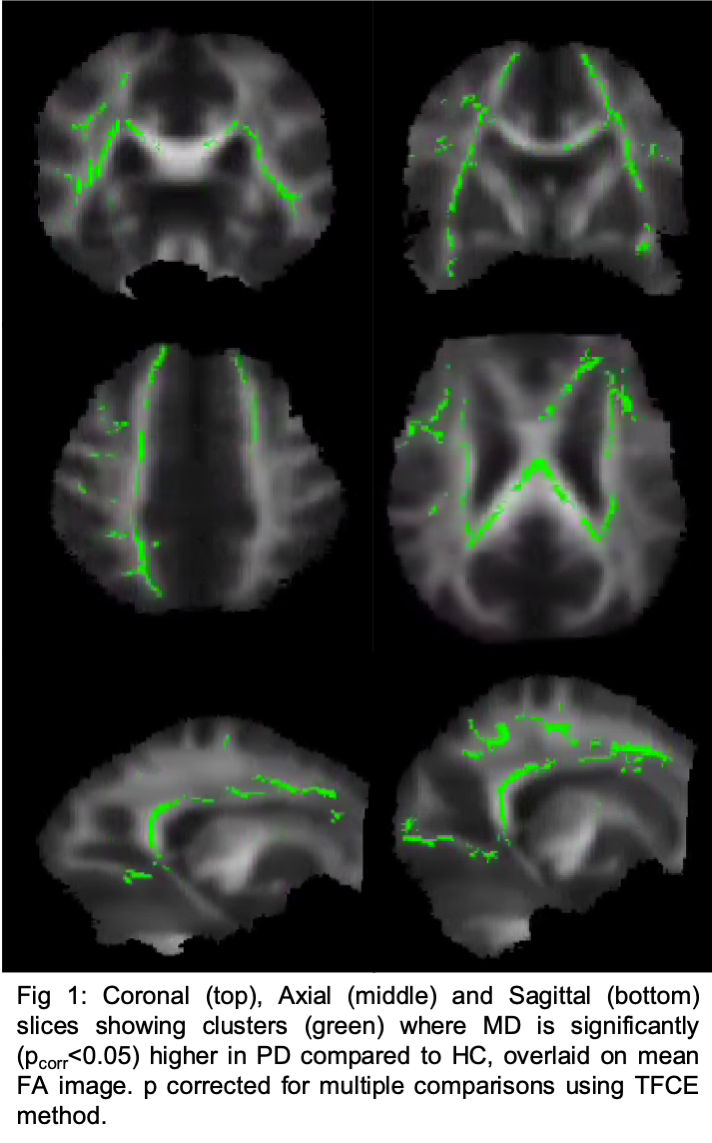
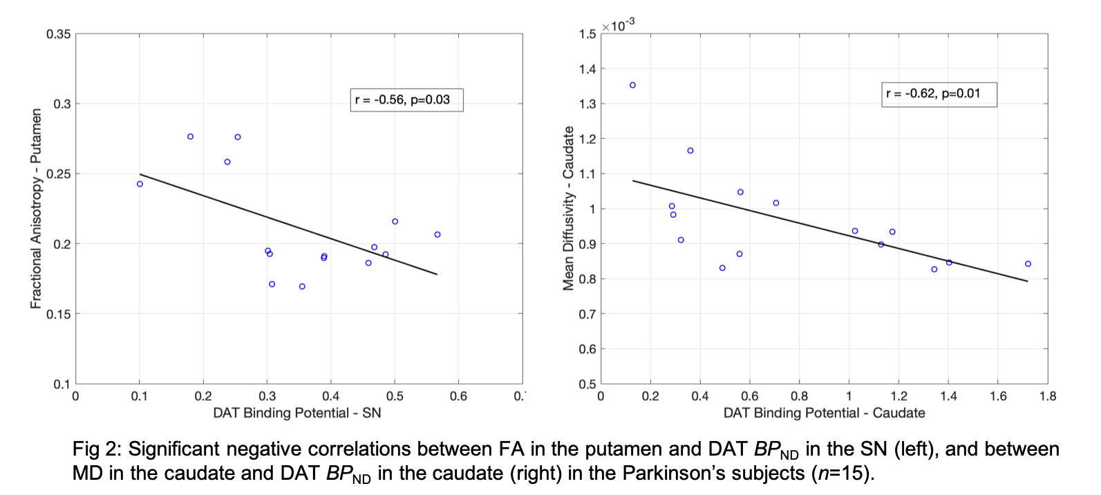
Results: Significantly higher MD (no change in FA) was observed in multiple WM tracts in PD subjects in TBSS analyses (Fig1). Regionally, FA was elevated in the putamen (11%, p=0.01) and SN (7.4%, p=0.07) of PD subjects. MD was higher in several cortical areas (Table1), particularly in the parietal and occipital cortex, precentral and postcentral gyri (~10% each, p<0.01). DTI outcomes were not significantly associated with MDS-UPDRS Part III. DAT availability was significantly lower in the SN, caudate and putamen in PD (range: [33-66]% lower, p < 0.01, Table2) and BPND in the putamen was negatively associated with MDS-UPDRS Part III (r=-0.58,p=0.03). In PD subjects, DAT BPND in SN correlated negatively with FA in the putamen (r=-0.56,p=0.03,Fig2). DAT BPND was also negatively associated with MD (r=-0.67,p=0.01,Fig2) in the caudate.
Conclusion: Globally increased MD and higher FA in the putamen could be characteristic features of PD-induced microstructure alterations.
To cite this abstract in AMA style:
P. Honhar, S. Tinaz, M. Dias, R. Comley, S. Finnema, R. Carson, D. Scheinost, D. Matuskey. Multimodal neuroimaging of Parkinson’s disease with diffusion tensor imaging (DTI) and dopamine transporter (DAT) positron emission tomography [abstract]. Mov Disord. 2023; 38 (suppl 1). https://www.mdsabstracts.org/abstract/multimodal-neuroimaging-of-parkinsons-disease-with-diffusion-tensor-imaging-dti-and-dopamine-transporter-dat-positron-emission-tomography/. Accessed December 23, 2025.« Back to 2023 International Congress
MDS Abstracts - https://www.mdsabstracts.org/abstract/multimodal-neuroimaging-of-parkinsons-disease-with-diffusion-tensor-imaging-dti-and-dopamine-transporter-dat-positron-emission-tomography/


