Category: Parkinson's Disease: Genetics
Objective: To explore whether autosomal recessive forms of monogenic PD (mPD) might lead to cortical and white matter (WM) abnormalities in addition to dopaminergic denervation.
Background: Previous studies have shown cortical and WM changes in idiopathic PD (iPD). But, there is limited data about cortex and WM pathology in mPD which is considered to be a “brain-first” subtype. In this regard, multimodal imaging may provide valuable insights for understanding mPD pathophysiology.
Method: 40 patients (17 Parkin, 3 DJ-1 mutation carrying mPD, 20 iPD shown to have no known PD mutations) were included. Heart-to-mediastinum, striatum/substantia nigra (SN)-to-occipital ratio obtained from cardiac and brain FDOPA PET/CT were compared with controls matched for age and gender. FDG PET images were evaluated visually. Diffusion Tensor Imaging was compared between PD groups using Tract Based Spatial Statistics (TBSS).
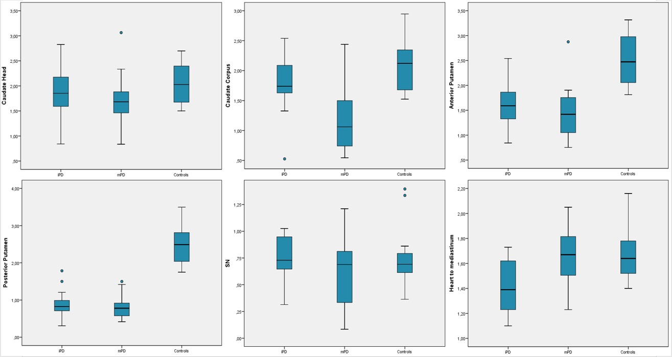
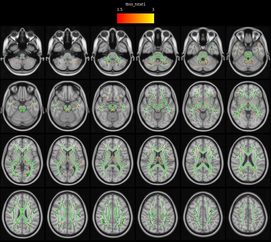
Results: The mean age of mPD was younger (43±9 vs 52±8 p:0.003), the mean age of disease onset was earlier (29±8 vs 46±8 p<0.001), disease duration was longer (14±7 vs 5±2 y, p<0.001) and H-Y stage was higher (1.9±0.5 vs 1.2±0.6 p:0.001) compared to iPD. mPD group showed a significant decrease in FDOPA uptake in caudate corpus compared to iPD and controls (n=13). A decrease in FDOPA uptake in SN was again more prominent in mPD without reaching statistical significance [figure1]. Myocardial FDOPA uptake in the mPD group (n=19, 1.64±0.2) was similar to controls (n=10, 1.66±0.2) whereas it was significantly reduced in iPD (n=20, 1.41±0.2 p:0.003). The cortical FDG uptake pattern was similar in mPD (n=19) and iPD (n=17). Hypometabolism was more conspicuous in the mesial prefrontal, inferior parietal and temporal cortices in both groups. TBSS analysis revealed a decrease in fractional anisotropy in WM areas, such as mesial temporal, bilateral cingulum and fornices, internal and external capsules, and superior longitudinal gyrus in mPD (n=17) compared to iPD (n=15) [figure2].
Conclusion: Compared to iPD, mPD patients showed an additional decrease in FDOPA uptake in caudate corpus and a tendency to decrease in SN, suggesting a more severe and widespread loss of dopaminergic neurons, despite a milder and slower disease process. The presence of metabolic and structural changes in the cortex and WM together with preserved myocardial innervation supports the idea of the brain-first progression of mPD.
To cite this abstract in AMA style:
B. Soydas-Turan, G. Yalcin-Cakmakli, E. Yetim, B. Volkan-Salanci, E. Lay-Ergun, K. Karli-Oguz, B. Elibol. Multimodal imaging analysis of autosomal recessive Parkinson’s disease. [abstract]. Mov Disord. 2023; 38 (suppl 1). https://www.mdsabstracts.org/abstract/multimodal-imaging-analysis-of-autosomal-recessive-parkinsons-disease/. Accessed April 17, 2025.« Back to 2023 International Congress
MDS Abstracts - https://www.mdsabstracts.org/abstract/multimodal-imaging-analysis-of-autosomal-recessive-parkinsons-disease/


