Category: Parkinson's Disease: Neuroimaging
Objective: To study nigrostriatal changes with a multifactorial neuroimaging approach in order to further characterize early abnormalities in Parkinson’s Disease (PD).
Background: Imaging techniques allow studying in-vivo the neurodegenerative mechanisms that relate to PD neuropathology. Alterations in several biomarkers such as R2* (iron concentration), fractional free-water volume (FW, microstructural integrity) and FDOPA uptake rate (Ki, dopaminergic activity) have already been described at PD onset1-3. However, a multimodal comprehensive characterization of these biomarkers in the early clinical stage of PD is still lacking.
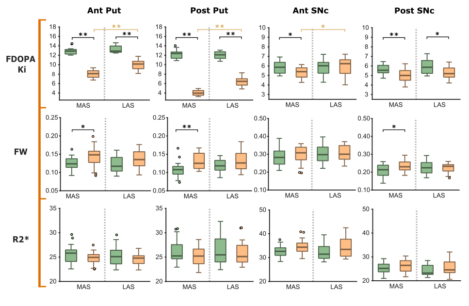
Method: 30 de novo PD (dN-PD) patients and 20 healthy subjects (HS) underwent PET and MRI studies no later than 12 months from the clinical diagnosis. From these sessions Ki, FW and R2* maps were reconstructed and assessed within putamen and substantia nigra pars compacta (SNc), which were partitioned into anterior and posterior divisions. Single hemibrain data was sorted according to predominance of motor signs (dN-PD) or hand dominance (HS), i.e., more and less affected sides (MAS/LAS) or dominant and non-dominant sides (DS/nDS). Non-parametric Mann-Whitney’s U tests were applied to study the inter-hemispheric asymmetries within groups and the inter-group differences caused by PD onset (MAS vs DS and LAS vs nDS).
Results: In dN-PD, Ki showed inter-hemispheric asymmetries in the putaminal and the anterior SNc, showing the MAS lower values (P<0.01). No asymmetries were displayed in any region for FW or R2*. There were significant reductions for Ki in the posterior (67/47% in MAS/LAS, P<0.01) and in the anterior (37/25% in MAS/LAS, P<0.01) putaminal divisions. In the posterior SNc a bilateral reduction of Ki was found (~11%, P<0.05), while in the anterior SNc only the MAS displayed a significant difference (9.5%, P<0.05). FW was increased in the MAS of both anterior and posterior putaminal divisions (~11%, P<0.05) and in the posterior SNc (11%, P < 0.05). No significant differences were observed in R2*.
Conclusion: Early PD involves both dopaminergic fibre loss and microstructural alterations in the nigrostriatal system. Dopaminergic depletion was clearly asymmetric in contrast to the microstructural changes, in keeping with the notion that dopaminergic denervation precedes cellular death. Iron disruption may develop in later disease stages.
References: 1. A. Brück, S. Aalto, E. Rauhala, J. Bergman, R. Marttila, and J. O. Rinne, “A follow-up study on 6-[18F]Fluoro-L-dopa uptake in early Parkinson’s disease shows nonlinear progression in the putamen,” Movement Disorders, vol. 24, no. 7, pp. 1009–1015, 2009.
2. G. Du et al., “Combined R2* and Diffusion Tensor Imaging Changes in the Substantia Nigra in Parkinson’s Disease,” Movement Disorders, vol. 26, no. 9, 2011.
3. E. Ofori et al., “Increased free water in the substantia nigra of Parkinson’s disease: A single-site and multi-site study,” Neurobiology of Aging, vol. 36, no. 2, pp. 1097–1104, 2015
To cite this abstract in AMA style:
M. López-Aguirre, M. Matarazzo, J. Blesa, A. Sánchez-Ferro, M. Hm Monje, J. Obeso, J. Pineda-Pardo. Multimodal assessment of nigrostriatal degeneration in de novo Parkinson’s disease [abstract]. Mov Disord. 2022; 37 (suppl 2). https://www.mdsabstracts.org/abstract/multimodal-assessment-of-nigrostriatal-degeneration-in-de-novo-parkinsons-disease/. Accessed April 18, 2025.« Back to 2022 International Congress
MDS Abstracts - https://www.mdsabstracts.org/abstract/multimodal-assessment-of-nigrostriatal-degeneration-in-de-novo-parkinsons-disease/

