Session Information
Date: Thursday, June 8, 2017
Session Title: Parkinson's Disease: Neuroimaging And Neurophysiology
Session Time: 1:15pm-2:45pm
Location: Exhibit Hall C
Objective: To validate Parkinson’s Disease-related network topography characterized with resting-state functional MRI (rs-fMRI) in two multicenter based cohorts of idiopathic Parkinson’s disease (IPD) patients.
Background: We recently developed a novel method, using independent component analysis in conjunction with bootstrap resampling to characterize specific network topographies associated with IPD (Vo et al, 2016). The functional PD-related pattern (fPDRP) discriminated normal controls (NC) from IPD patients and showed a similar topography compared to the previously characterized pattern identified using metabolic PET imaging.
Methods: We studied two independent cohorts of IPD patients of three different sites (Center(C) 1 Northshore University Hospital, C2 University of Stanford & Pennsylvania) and NC subjects with rs-fMRI in a medication-free (off) state. Subject scores and temporal dynamics were estimated for prospective cases with dual regression using the spatial maps generated from the derivation set (Vo et al, 2016). Individual values were z-scored with respect to corresponding values from the healthy control group. Motor symptoms were assessed in all IPD subjects using the Unified Parkinson’s Disease Rating Scale (UPDRS).
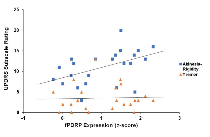
Results: We scanned 30 IPD patients, divided into two different cohorts (C1 n=19, C2 n=11), and 18 age- and gender matched healthy controls. Expression values for fPDRP were significantly elevated in both testing cohorts (C1 p<0.001; C2 p=0.02) compared to normal controls (Figure 1). Subject scores correlated with subscale ratings for akinesia-rigidity (r=0.47, p<0.002) but not tremor (r=0.14, p=0.51) in this multicenter testing cohort (Figure 2).
Conclusions: Our findings reveal that fPDRP represents a replicable imaging marker across independent multicenter cohorts of IPD patients. These results provide further support for the stability of the fPDRP topography as well as the consistency of its relationship to motor symptoms across different patient populations.
References: Vo A, Sako W, Fujita K, et al (2016) Parkinson’s disease-related network topographies characterized with resting state functional MRI. Hum Brain Mapp.
To cite this abstract in AMA style:
K. Schindlbeck, A. Vo, Y. Ma, S. Peng, K. Poston, H. Hurtig, N. Dahodwala, D. Eidelberg. Multicenter Validation of Disease- related Parkinson’s disease pattern with Resting State functional MRI [abstract]. Mov Disord. 2017; 32 (suppl 2). https://www.mdsabstracts.org/abstract/multicenter-validation-of-disease-related-parkinsons-disease-pattern-with-resting-state-functional-mri/. Accessed April 18, 2025.« Back to 2017 International Congress
MDS Abstracts - https://www.mdsabstracts.org/abstract/multicenter-validation-of-disease-related-parkinsons-disease-pattern-with-resting-state-functional-mri/


