Category: Parkinson's Disease: Neuroimaging
Objective: To compare MRI sings and volumetric measurements in Parkinson disease (PD), Progressive Supranuclear Palsy (PSP), and Multiple System Atrophy (MSA) and their relationship with clinical motor scale.
Background: MRI signs and volumetric measurements have been proposed to facilitate the differential diagnosis between parkinsonian syndromes and helps to understand the pathophsyiology underlining these syndromes.
Method: Sociodemographic, clinical, and MRI characteristics were obtained from 240 consecutive patients from a Mexican Movements Disorders clinic. They were classified according to clinical diagnosis as PD=170; PSP=12; MSA=13; as well as, a control group (n=27) used for comparisons.
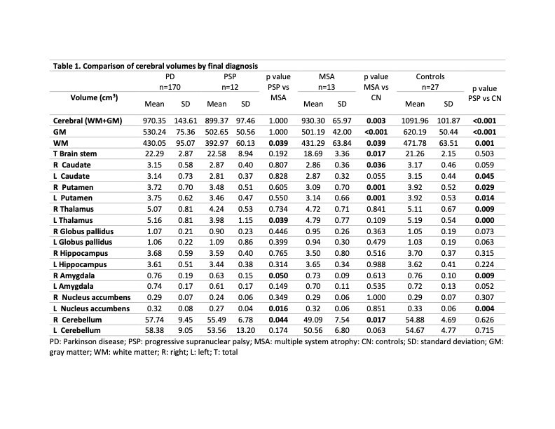
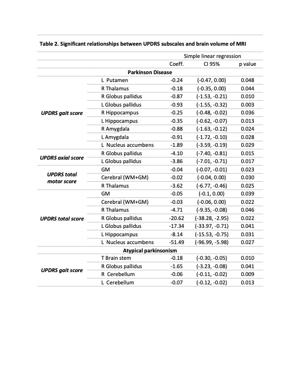
Results: Significant differences were found between groups in age at evaluation, age at onset, UPDRS motor and total scores, and cognitive assessments. Atypical parkinsonian (AP) cluster were older at initial evaluation, as well as, at symptoms onset. They showed higher UPDRS scores and lower cognitive performance. Higher percentage of AP showed imaging signs (i.e.: Hummingbird sign/Morning glory signs). PD showed higher brain volumes than PSP, except for the brainstem and left Globos Pallidus (GP). Differences in volumetric measures were significantly higher for White Matter (WM), thalamus, GP, amygdala, and the left nucleus accumbens in PD as well. Nevertheless, PD group had lower WM and left hippocampal volumes compared to MSA. Compared to controls, PD group had significantly lower total brain, Grey Matter (GM), and WM volumes and the left cerebellum volume was higher than the control group [table 1]. In PD, significant relationships were found between the UPDRS total score and the GM, GM+WM, right thalamus, GP, left hippocampus, and left nucleus accumbens volumes. In AP patients, UPDRS gait score showed a relationship with brainstem volume and cerebellum [table 2].
Conclusion: AP patients showed higher disease severity (UPDRS and cognitive scores), although disease duration was similar than PD. In PD, total brain and basal ganglia volumes showed a significant relationship with motor scales. For AP, brain stem and cerebellum measurements are related with motor characteristics. Imaging features are not only useful in differential diagnosis; but also, in understanding the pathophysiological features of PD and other AP syndromes.
To cite this abstract in AMA style:
H. Martinez-Hernandez, D. Lopez-Mena, B. Candela-Solano, A. Medina-Islas, L. Alvarez, I. Acosta. Multi-modal MRI features and their relationship with clinical scales in Parkinson disease and Atypical Parkinsonian Syndromes [abstract]. Mov Disord. 2022; 37 (suppl 2). https://www.mdsabstracts.org/abstract/multi-modal-mri-features-and-their-relationship-with-clinical-scales-in-parkinson-disease-and-atypical-parkinsonian-syndromes/. Accessed February 1, 2026.« Back to 2022 International Congress
MDS Abstracts - https://www.mdsabstracts.org/abstract/multi-modal-mri-features-and-their-relationship-with-clinical-scales-in-parkinson-disease-and-atypical-parkinsonian-syndromes/


