Category: Parkinson's Disease: Neuroimaging
Objective: We aimed to investigate underlying modifications of MRI functional connectivity between dysarthric and non-dysarthric patients with Parkinson’s disease and healthy controls.
Background: Altered speech production is a common symptom in Parkinson’s disease (PD), is not responsive to dopaminergic therapy and can be
worsened by deep brain stimulation. A better comprehension of the functional connectivity of dysarthric and non-dysarthric PD patients may lead to
new therapeutic approaches, and to the possibility to determine if a PD patient is likely to become dysarthric or not.
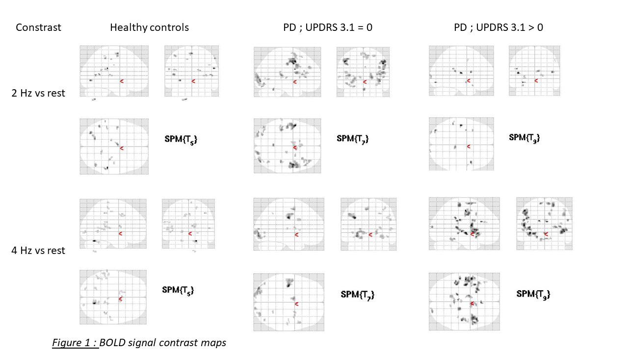
Method: We studied 24 PD patients in the ON dopaminergic conditions and 13 healthy controls (HC). Dysarthria was defined by a score ≥1 on UPDRS item 3.1. An overt speech task (repetition of /pa/ syllable at a frequency of 2 or 4 Hz [1]) was conducted during the imaging, and the blood-oxygen-level dependent (BOLD) signal was measured. Then, we generated for each group two activation maps, one for contrast 2 Hz vs rest, and a second for 4 Hz vs rest. Then we compared these maps between groups.
Results: Due to movements artifacts, contrast maps were generated from 6 HC, 8 non-dysarthric PD and 10 dysarthric PD. With correction for multiple comparisons, there were no significant differences (pcorr < 0.05) between groups for the 2 prespecified contrasts. Thus, for the 2Hz vs rest contrast, there is a trend (punc< 0.001, cluster size > 10 voxels) for a more important BOLD signal in speech production-related areas in non-dysarthric PD patients, compared to HC and dysarthric PD patients. For the 4 Hz vs rest contrast, there is no major BOLD signal differences between HC and non-dysarthric PD patients, but there is a trend for a more important BOLD signal in speech production-related areas in dysarthric patients [figure1].
Conclusion: Our results suggest that non-dysarthric PD patients may present an early compensatory cerebral overactivation, which may be delayed for dysarthric patients resulting in altered speech production. Further explorations could investigate correlations between BOLD signal and a continuous dysarthria score, for speech tasks and resting-state functional MRI.
References: [1] Moreau, Caroline, et al. « Oral Festination in Parkinson’s Disease: Biomechanical Analysis and Correlation with Festination and Freezing of Gait ». Movement Disorders, vol. 22, no 10, 2007, p. 1503‑06. Wiley Online Library, doi:10.1002/mds.21549.
To cite this abstract in AMA style:
J.P Niguet, R. Viard, T. Guichoux, A. Basirat, L. Defebvre, G. Kuchcinski, D. Devos, C. Moreau. MRI functional connectivity analysis for speech impairment in Parkinson’s disease [abstract]. Mov Disord. 2020; 35 (suppl 1). https://www.mdsabstracts.org/abstract/mri-functional-connectivity-analysis-for-speech-impairment-in-parkinsons-disease/. Accessed April 21, 2025.« Back to MDS Virtual Congress 2020
MDS Abstracts - https://www.mdsabstracts.org/abstract/mri-functional-connectivity-analysis-for-speech-impairment-in-parkinsons-disease/

