Category: Parkinson's Disease: Neuroimaging
Objective: To develop a motion-insensitive high-resolution susceptibility weighted imaging (SWI) sequence for evaluating nigrosome-1 in substantia nigra.
Background: In Parkinson’s disease (PD) increased amounts of iron are seen in nigrosome-1. This can be detected using SWI as a loss of the ‘swallow tail sign’. However, MRI is sensitive to motion, which can degrade image quality, especially in small structures such as nigrosome-1. Furthermore, an advantage of SWI for iron detection is that it uses both magnitude and phase, but motion-induced phase changes are difficult to adjust for. Consequently, non-diagnostic SWI images are common due to motion artifacts. In a well-conducted study, 16% of participants were excluded, and of the excluded 73% had PD [1].
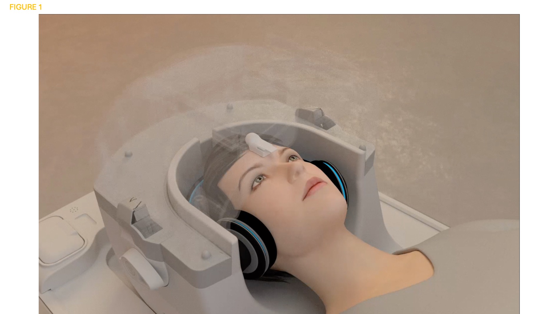
Method: We further developed a 2D SWI sequence using interleaved snapshot sampling [2]. It collects all data for each slice and NEX (number of excitations) within a minimum time frame, thereby lessening motion-induced phase changes. A free induction (FID) navigator was also incorporated [3], which corrects for breathing-induced phase changes. For prospective motion correction we used an in-house developed wireless RF triggered acquisition device mounted on the forehead (Figure 1), which with high temporal speed and accuracy adjusts for submillimeter motion [4]. Finally, noise reduction was performed using optimized blockwise nonlocal means [5]. Spatial resolution was 0.7 x 0.7 x 1 mm (8.16 minutes). For comparison a high-resolution 3D SWI was performed, 0.34 x 0.34 x 2 mm (2.56 minutes), also using prospective motion correction [4]. A PD patient was evaluated with both sequences, once with ‘no motion’ and once rotating the head left-right (LR) continuously during the scan. For reference, a healthy participant was evaluated with the 2D snapshot SWI sequence and ‘no motion’. A 3T scanner was used.
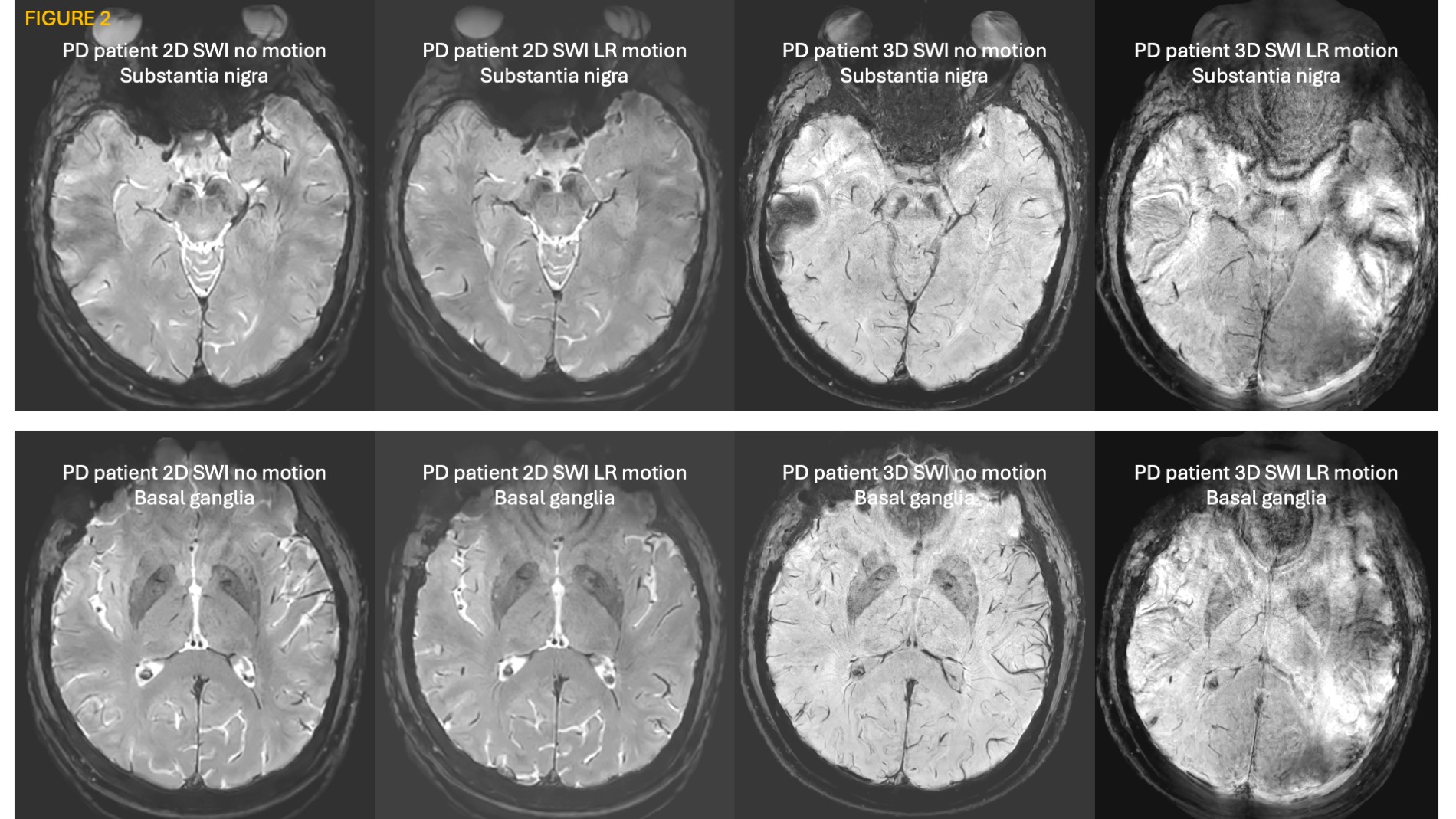
Results: Figure 2 shows good quality for 2D snapshot SWI, both in scans with ‘no motion’ and LR motion, while 3D SWI is non-diagnostic for LR motion, showing the fundamental effect of snapshot sampling in reducing motion-induced phase changes. PD patient with loss of ‘swallow tail sign’.
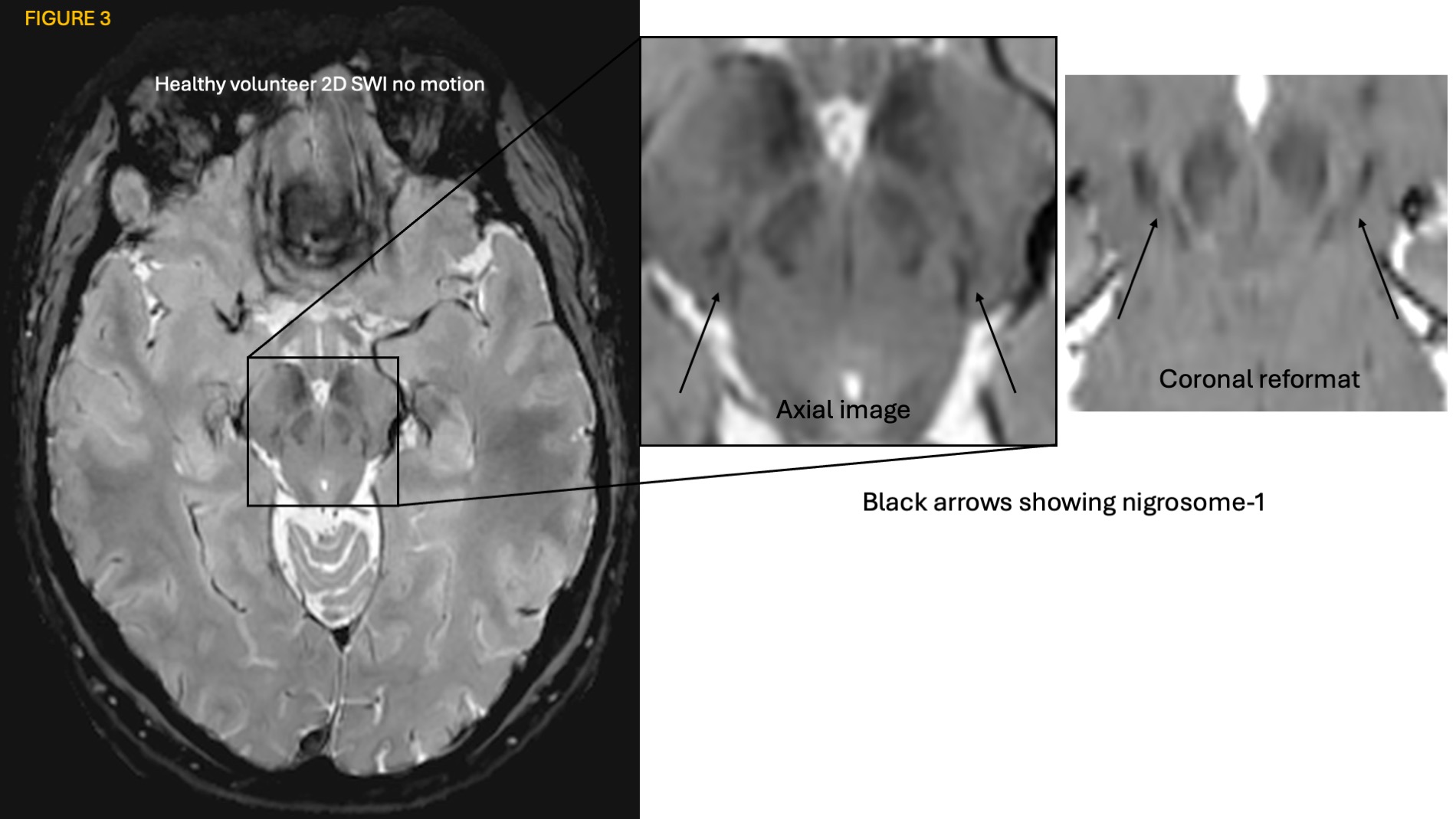
Figure 3 shows good quality when reformatting images in a coronal view. Healthy participant having the ‘swallow tail sign’.
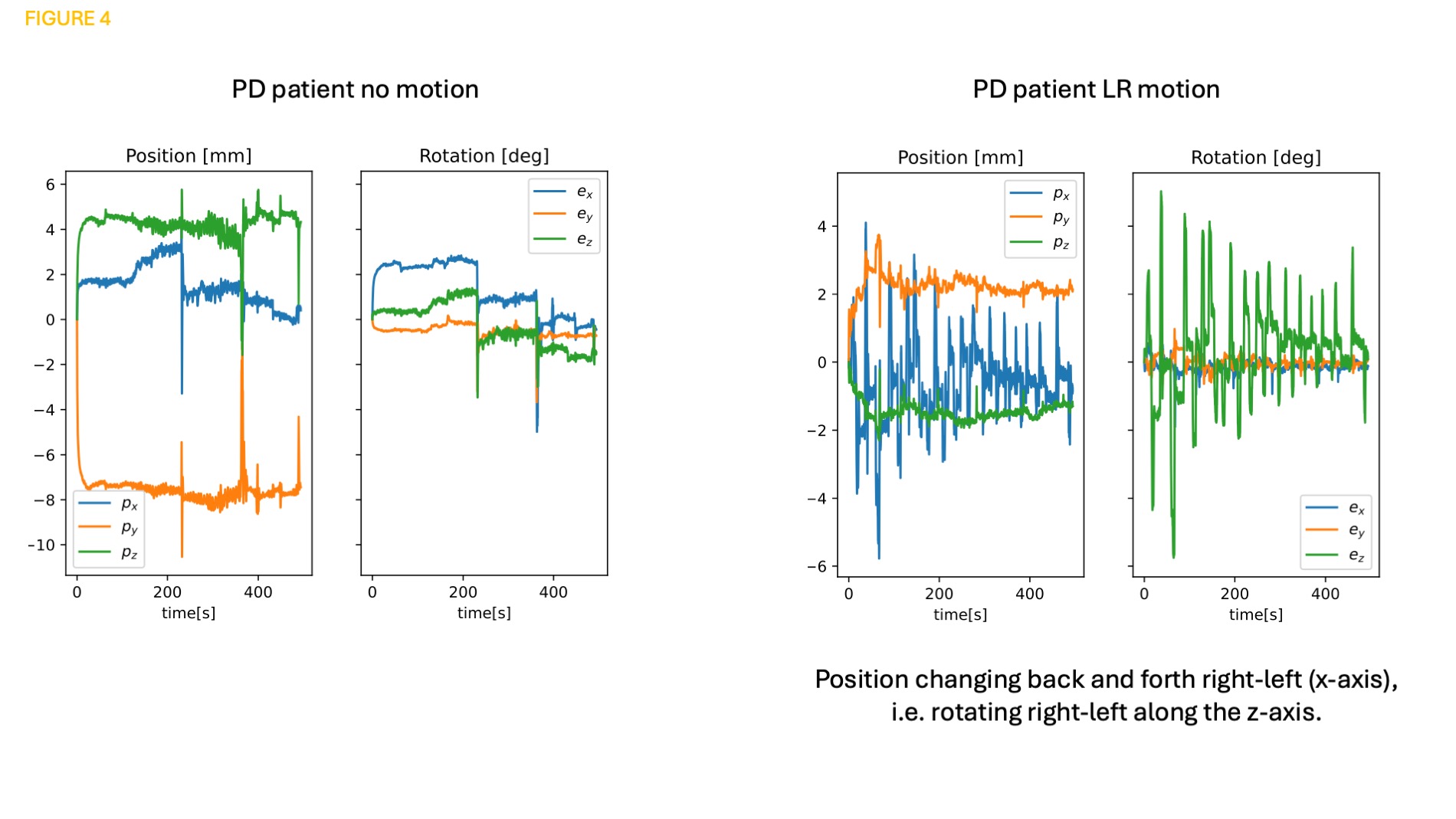
Figure 4 shows motion logs for the PD patient during the 2D snapshot SWI scans.
Conclusion: Motion robust high-resolution 2D SWI MRI is possible for evaluating nigrosome-1.
Figure 1
Figure 2
Figure 3
Figure 4
References: 1. Haller, S.; Davidsson, A.; Tisell, A.; Ochoa-Figueroa, M.; Georgiopoulos, C. MRI of nigrosome-1: A potential triage tool for patients with suspected parkinsonism. J Neuroimaging 2022, 32 (2), 273-278.
2. Berglund, J.; Sprenger, T.; van Niekerk, A.; Rydén, H.; Avventi, E.; Norbeck, O.; Skare, S. Motion-insensitive susceptibility weighted imaging. Magn Reson Med 2021, 86 (4), 1970-1982.
3. Pfeuffer J, Van de Moortele PF, Ugurbil K, Hu X, Glover GH. Correction of Physiologically Induced Global Off-Resonance Effects in Dynamic Echo-Planar and Spiral Functional Imaging. Magnetic Resonance in Medicine. 2002;47(2):344–353.
4. van Niekerk, A.; Berglund, J.; Sprenger, T.; Norbeck, O.; Avventi, E.; Rydén, H.; Skare, S. Control of a wireless sensor using the pulse sequence for prospective motion correction in brain MRI. Magn Reson Med 2022, 87 (2), 1046-1061.
5. Coupe P, Yger P, Prima S, Hellier P, Kervrann C, Barillot C. An optimized blockwise nonlocal means denoising filter for 3-D magnetic resonance images. IEEE Trans Med Imaging. 2008;27(4):425–441.
To cite this abstract in AMA style:
M. Skorpil, O. Norbeck, P. Svenningsson, S. Skare, A. van Niekerk. Motion-robust high-resolution imaging of nigrosome-1 in Parkinson’s disease [abstract]. Mov Disord. 2024; 39 (suppl 1). https://www.mdsabstracts.org/abstract/motion-robust-high-resolution-imaging-of-nigrosome-1-in-parkinsons-disease/. Accessed April 20, 2025.« Back to 2024 International Congress
MDS Abstracts - https://www.mdsabstracts.org/abstract/motion-robust-high-resolution-imaging-of-nigrosome-1-in-parkinsons-disease/


