Category: Tics/Stereotypies
Objective: Our objective is to identify the fiber tracts modulated during deep brain stimulation (DBS) of the globus pallidus internus (GPi) and identify those associated with tic improvement.
Background: Recent studies suggest that tic improvement following DBS for TS may rely on modulation of structural networks rather than specific anatomical regions [1,2]. It is unknown which specific fiber tracts should be modulated in order to most effectively improve tics.
Method: Retrospective imaging, longitudinal Yale Global Tic Severity Scale (YGTSS) total scores, and stimulation settings were acquired for TS patients who underwent bilateral GPi DBS. Computational models with patient-specific stimulation settings were used to estimate the activation of fiber tracts surrounding the GPi drawn by expert neuroanatomists [3]. The modeled tracts included the pallidothalamic tracts [lenticular fasciculus (LF), ansa lenticularis (AL)], tracts connecting the globus pallidus externus and the subthalamic nucleus (GPe-to-STN), and internal capsule (IC) tracts projecting to prefrontal cortex (PFC), premotor cortex (PMC), and motor cortex (MC). Linear mixed effects (LME) models were used to assess if the percent activation of the tracts was associated with improvement in YGTSS scores while accounting for time point, baseline score, and repeated outcome measures for each patient.
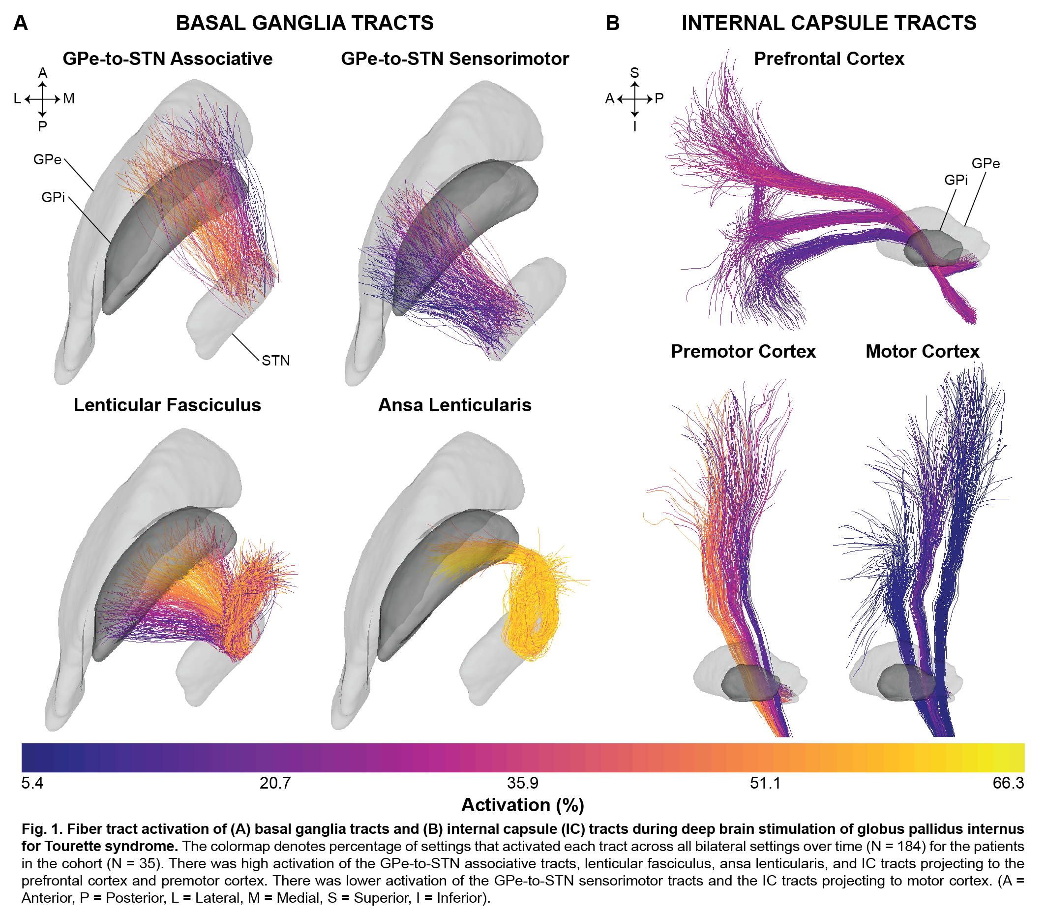
Results: Heatmaps of the percentage of bilateral stimulation settings (N=184) across all patients (N=35) that activated each fiber tract are shown in Fig. 1. The pallidothalamic tracts (LF and AL), GPe-to-STN associative tracts, and the IC tracts projecting to PFC and PMC showed higher activation, while there was low activation of the GPe-to-STN sensorimotor tracts and the IC tracts projecting to MC. The LME models showed that percent activation of the GPe-to-STN associative tracts (b=0.37, p<0.001) and the AL (b=0.32, p<0.001) was significantly associated with improvement in YGTSS total scores.[figure1]
Conclusion: Tic improvement following DBS of the GPi may depend on modulation of the GPe-to-STN associative tracts and the AL. The results provide insight into the potential mechanisms of tic improvement and could be used to refine stimulation targets. In the future we will develop LME models of combined tract activation to predict clinical outcomes and optimize stimulation settings in de novo TS DBS patients.
References: [1] Johnson KA, Fletcher PT, Servello D, et al. Image-based analysis and long-term clinical outcomes of deep brain stimulation for Tourette syndrome: a multisite study. J Neurol Neurosurg Psychiatry 2019;90:1078–90. doi:10.1136/jnnp-2019-320379. [2] Johnson KA, Duffley G, Anderson DN, et al. Structural Connectivity Predicts Clinical Outcomes of Deep Brain Stimulation for Tourette Syndrome. In Review. [3] Petersen M V, Mlakar J, Haber SN, et al. Holographic Reconstruction of Axonal Pathways in the Human Brain. Neuron 2019;104:1056-1064.e3. doi:10.1016/j.neuron.2019.09.030.
To cite this abstract in AMA style:
K. Johnson, T. Foltynie, M. Hariz, L. Zrinzo, E. Joyce, Z. Kefalopoulou, P. Limousin, H. Akram, D. Servello, A. Bona, M. Porta, J.G Zhang, F.G Meng, Z. Ling, X. Xu, X. Yu, A. Leentjens, A. Smeets, L. Ackermans, L. Almeida, A. Gunduz, W. Hu, K. Foote, M. Okun, C. Butson. Modulation of Basal Ganglia and Internal Capsule Fiber Tracts During Deep Brain Stimulation for Tourette Syndrome [abstract]. Mov Disord. 2020; 35 (suppl 1). https://www.mdsabstracts.org/abstract/modulation-of-basal-ganglia-and-internal-capsule-fiber-tracts-during-deep-brain-stimulation-for-tourette-syndrome/. Accessed December 18, 2025.« Back to MDS Virtual Congress 2020
MDS Abstracts - https://www.mdsabstracts.org/abstract/modulation-of-basal-ganglia-and-internal-capsule-fiber-tracts-during-deep-brain-stimulation-for-tourette-syndrome/

