Session Information
Date: Wednesday, September 25, 2019
Session Title: Neuroimaging
Session Time: 1:15pm-2:45pm
Location: Les Muses Terrace, Level 3
Objective: The aim of this study was to apply diffusion tensor imaging (DTI) to evaluate atrophy of the Substantia Nigra pars compacta (SNc), the pathological hallmark of Parkinson’s disease (PD), as a marker of disease severity.
Background: Significant variability exists in the reported DTI metrics of the SNc in PD due to variability in SNc localization and segmentation techniques. An accurate and standardised delineation of the SNc will improve accuracy and hence the potential of DTI metrics as a biomarker of PD severity.
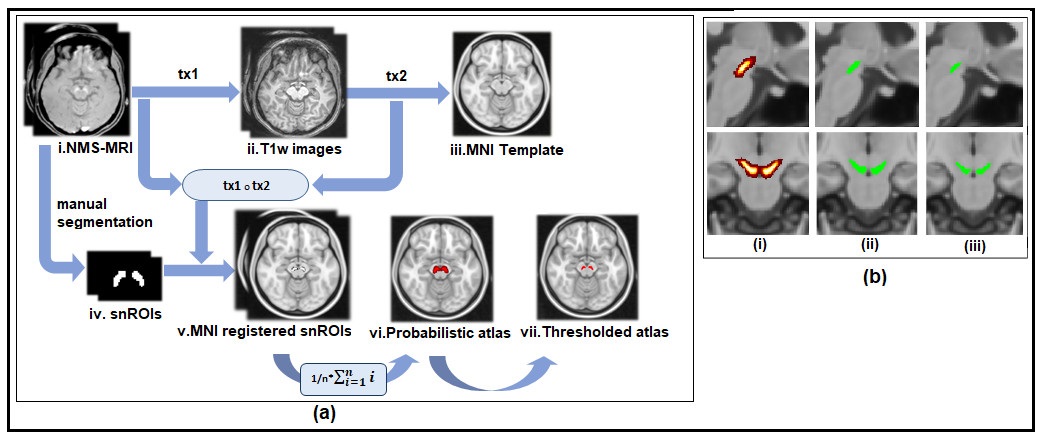
Method: DTI images of 86 healthy controls (HC) and 158 denovo PD patients were used from the PPMI database. A probabilistic atlas of SNc was created using different set of 27 controls, scanned on a 3T Philips Achieva scanner. A neuromelanin sensitive MRI (NM-MRI) was acquired using TR/TE: 26/2.2ms, FOV: 180x180x50mm, voxel size: 0.9×0.9x1mm, 50 slices, along with a T1w image. SNc atlas was constructed in MNI space as shown in [Figure1]. DTI images were noise filtered, corrected for eddy currents and head motion and were skull stripped. DTI maps of fractional anisotropy (FA), axial, mean and radial diffusivity (AD,MD,RD) were computed using ‘dtifit’ from FSL5.0.9. and were registered to MNI spaced FMRIB58 image. Diffusion measures of SN were extracted by thresholding atlas at 0.5 probability. MANCOVA model was applied on diffusion measures of HC and PD groups using age and gender as covariates. Pearson correlation was done between FA,AD,MD,RD and clinical scores (UPDRS III (Off), H&Y) of all age and gender corrected PD patients. An independent t-test was done between diffusion measures of HC and PD patients with a UPDRS score>50 percentile (PDhigh) to assess the micro structural changes occurring with disease severity.
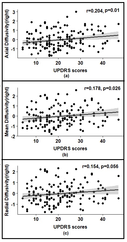
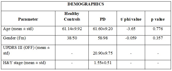
Results: Detailed demographic information of all subjects is shown in [Table1]. No significant differences in diffusion measures were seen between HC and PD. Only UPDRS scores showed a significant positive correlation with AD, MD and RD of right SNc as shown in [Figure2]. The AD (p=0.026) and MD (p=0.059) of right SNc was found to be significantly higher in PDhigh as compared to HC group.
Conclusion: High AD, MD and RD was associated with higher disease severity in PD, indicating abnormal axonal density and an increase in demyelination of SNc. Our findings strongly suggest that AD, MD and RD could be used as potential biomarkers for evaluating severity and disease progression in PD.
To cite this abstract in AMA style:
A. Safai, S. Rane, S. Prasad, J. Saini, P. Pal, M. Ingalhalikar. Micro structural Changes in Substantia Nigra are associated with Severity of Parkinson’s Disease. [abstract]. Mov Disord. 2019; 34 (suppl 2). https://www.mdsabstracts.org/abstract/micro-structural-changes-in-substantia-nigra-are-associated-with-severity-of-parkinsons-disease/. Accessed April 21, 2025.« Back to 2019 International Congress
MDS Abstracts - https://www.mdsabstracts.org/abstract/micro-structural-changes-in-substantia-nigra-are-associated-with-severity-of-parkinsons-disease/


