Session Information
Date: Wednesday, September 25, 2019
Session Title: Neuroimaging
Session Time: 1:15pm-2:45pm
Location: Les Muses Terrace, Level 3
Objective: The objective of this study is to identify and validate the metabolic PD-related pattern (PDRP) in de novo PD patients and to compare it to the original PDRP, identified in more advanced PD patients.
Background: Metabolic changes are observed in patients with Parkinson disease (PD) and known to increase with advancing disease stages. The PD-related metabolic pattern (PDRP) has been validated as an imaging biomarker candidate for the assessment of disease progression and new therapies in PD (Schindlbeck and Eidelberg, 2018). Little is known whether respective brain changes are already present in early stages of PD before the exposure to dopaminergic therapy (drug-naïve PD).
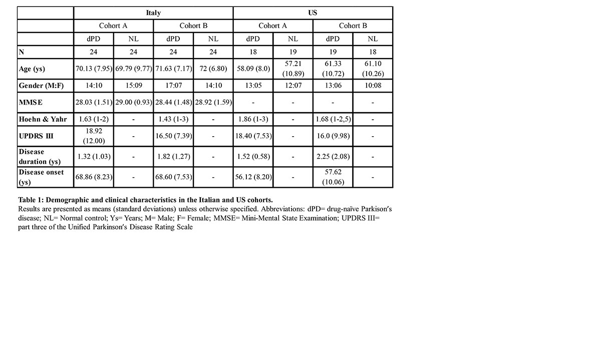
Method: We studied two independent cohorts of de novo diagnosed, drug-naïve patients with PD (dPD) and respective healthy control groups (NL) with clinical assessments and metabolic imaging with 18F-FDG-PET. The Italian (dPD/NL: n=48/48) and US (dPD/NL: n=37/37) cohorts were randomly divided into cohort A and B [Table 1]. Spatial covariance analysis in cohort A was used to identify dPD-related patterns (dPDRP) in the Italian and US cohorts. Cohort B was used to subsequently validate the dPDRP identified in cohort A. Expression levels of these patterns in dPD patients were compared to NL. The dPDRP networks were compared to the previously known PDRP using subject-score correlations and voxel-wise correlations for topographical overlap.
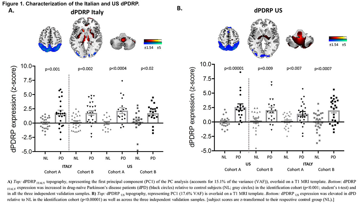
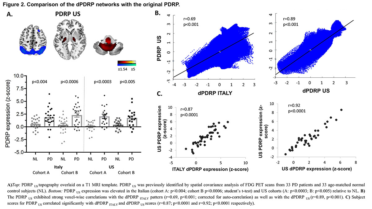
Results: dPDRP-Italy and dPDRP-US topographies were both characterized by relatively increased metabolic activity in the globus pallidum, putamen, thalamus, pons, cerebellum and sensorimotor cortex, and by reduced metabolic activity in the lateral cortex and parieto-occipital association regions [Fig.1]. Expression levels were significantly elevated in dPD patients compared to NL in the identification as well as validation cohorts [Fig.1]. Both dPDRPs exhibited strong topographic similarities with the original PDRP [Fig.2]. Expression values of the dPDRP Italy and the dPDRP US correlated strongly with subject scores of the original PDRP in the Italian and US cohorts.
Conclusion: Metabolic network abnormalities are already evident in de novo PD patients and significantly elevated relative to healthy control subjects. These findings are consistent with prior reports of PDRP elevations in the “preclinical” hemispheres of hemi-PD patients and in patients with REM-sleep behaviour disorder.
References: Schindlbeck KA, Eidelberg D (2018) Network imaging biomarkers: insights and clinical applications in Parkinson’s disease. Lancet Neurol 17:629–640. doi: 10.1016/S1474-4422(18)30169-8
To cite this abstract in AMA style:
O. Lucas-Jimenez, K. Schindlbeck, C. Tang, S. Morbelli, D. Arnaldi, M. Pardini, M. Pagani, F. Nobili, D. Eidelberg. Metabolic network abnormalities in drug-naive Parkinson’s disease [abstract]. Mov Disord. 2019; 34 (suppl 2). https://www.mdsabstracts.org/abstract/metabolic-network-abnormalities-in-drug-naive-parkinsons-disease/. Accessed April 21, 2025.« Back to 2019 International Congress
MDS Abstracts - https://www.mdsabstracts.org/abstract/metabolic-network-abnormalities-in-drug-naive-parkinsons-disease/


