Session Information
Date: Thursday, June 8, 2017
Session Title: Parkinson's Disease: Neuroimaging And Neurophysiology
Session Time: 1:15pm-2:45pm
Location: Exhibit Hall C
Objective: As part of a pioneer trial to investigate the use of transcranial magnetic resonance-guided focused ultrasound subthalamotomy (MRgFUS-STN) in Parkinson’s disease (PD), this study addresses the effects of sonication on resting-state cerebral glucose metabolism and examines the associations between lesions size and modulation of brain metabolism.
Background: Subthalamotomy is effective in alleviating motor symptoms in PD. Recently, MRgFUS has become a minimally-invasive tool aimed to precisely generate thermocoagulation in deep brain structures.
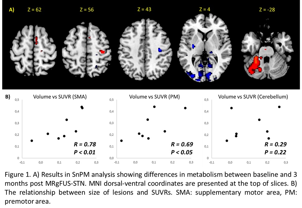
Methods: Eight PD patients receiving MRgFUS-STN underwent simultaneous FDG-PET/MRI at rest in off-medication, both previous and three months after procedure. MRI were segmented and normalized using the DARTEL method and volume of lesions were computed. PET were corrected for partial volume effect, normalized and smoothed using a 6-mm gaussian kernel. Standardized uptake ratios (SUVRs) were computed using pons as reference. We performed voxel-based statistical non-parametric mapping (SnPM) based upon 10.000 random permutations to identify significant changes in regional metabolism that occurred with MRgFUS-STN. ROI-based hypothesis-driven correlations between volume of lesions and SUVRs were performed for motor areas.
Results: All treatments were recognized by MRI. Average volume of lesions was 0.32 cc, involving the sensorimotor STN and dorsal white matter. SnPM analysis revealed a significant impact on brain metabolism characterized by increase in ipsilateral premotor (PM) and supplementary motor area (SMA), brainstem and contralateral cerebellum, and reductions in the ipsilateral primary motor area, striatum, thalamus, contralateral superior temporal and bilateral posterior putamen and posterior parietal/occipital regions. There was a significant correlation between volume of lesions and glucose metabolism in SMA and PM. (Figure 1).
Conclusions: MRgFUS-STN modulates the activity of PD-related metabolic brain networks. Contrary to classical approach using DBS, our data are consistent with deactivation of the inhibitory basal ganglia output and subsequent activation of premotor areas. Modulation of posterior areas suggest a complex inter-network interaction induced by the influence of lesion over fibre tracts crossing the subthalamic region. ROI-based analysis documents a relation between the size of the lesion and the amplitude of changes in metabolism.
To cite this abstract in AMA style:
R. Rodriguez-Rojas, R. Martinez-Fernandez, J. Pineda-Pardo, C. Sanchez-Catasus, M. del Alamo, J. Obeso. Metabolic changes following transcranial magnetic resonance-guided focused ultrasound subthalamotomy [abstract]. Mov Disord. 2017; 32 (suppl 2). https://www.mdsabstracts.org/abstract/metabolic-changes-following-transcranial-magnetic-resonance-guided-focused-ultrasound-subthalamotomy/. Accessed April 18, 2025.« Back to 2017 International Congress
MDS Abstracts - https://www.mdsabstracts.org/abstract/metabolic-changes-following-transcranial-magnetic-resonance-guided-focused-ultrasound-subthalamotomy/

