Category: Neuroimaging (Non-PD)
Objective: To describe the clinical manifestations and structural and functional neuroimaging findings in a patient with a left mesencephalic neuroglial cyst, resembling a PSP.
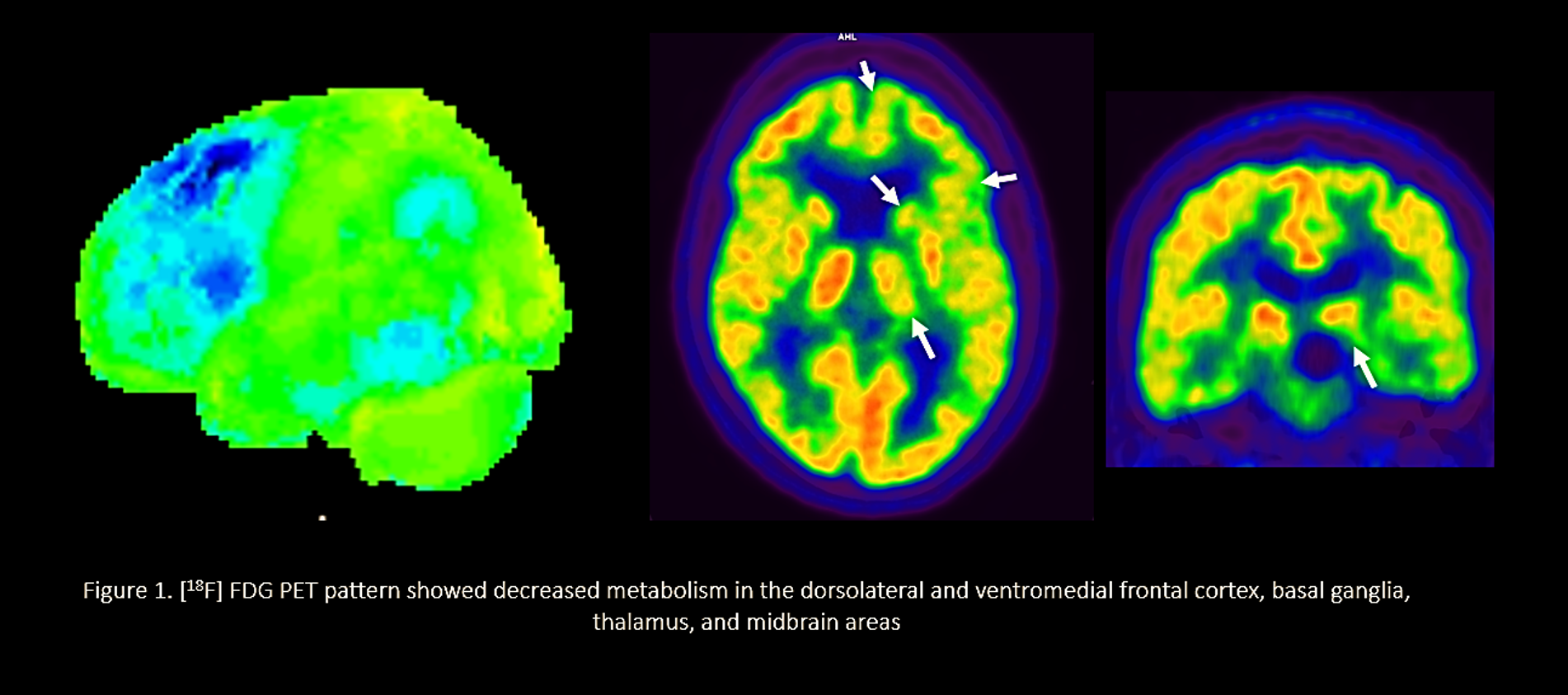
Background: PSP diagnosis is based on the presence of clinical manifestations, and abnormalities in MRI and [18F] dopa and [18F] FDG PET scans can serve as supporting criteria. The [18F] FDG PET pattern is characterized by decreased metabolism in the dorsolateral and ventromedial frontal cortex, basal ganglia, thalamus, and midbrain areas. Intraparenchymal cysts of the mesencephalon can produce manifestations of intracranial hypertension; however, parkinsonism and other movement disorders are very uncommon.
Method: Clinical case description and literature review.
Results: An 87-year-old woman with a two-year history of motor clumsiness in the right limbs and gait disturbance. Her past medical history included a gastrointestinal stromal tumor. A small left mesencephalic cyst had been diagnosed five years ago and was an incidental finding. At the examination, she exhibited dysarthria, lower right facial asymmetry, mild bimanual apraxia, and supranuclear vertical gaze palsy. Strength and muscle tone were normal. Right bradykinesia was observed. No tremor, rigidity, myoclonus, or sensory deficits. Plantar responses were flexor bilaterally. Her gait was unstable with short steps and decreased arm swing on her right-hand side. The cognitive assessment revealed mild dysexecutive cognitive impairment. A new brain MRI showed that the cyst had significantly increased, and was affecting the left side of the midbrain (13 mm in diameter). DTI tractography showed alteration in fractional anisotropy value; distortion of several tracts, and substantia nigra.
Two [18F] dopa PET scans performed 3 years apart did not show any alteration. However, [18F] FDG PET scan revealed bilaterally, dorsolateral, and dorsomedial frontal cortical hypometabolism with the left side more affected, also affecting the caudate nucleus, thalamus, and left midbrain.
Conclusion: Our patient presented a clinical phenotype with metabolic [18F] FDG PET pattern resembling PSP. We suggest that the mesencephalic lesion might produce modifications in cerebral metabolism, and give raise to the clinical phenotype observed in PSP patients. However, we cannot rule out the existence of an underlying degenerative process.
References: 1. Endo H, Fujimura M, Watanabe M, Tominaga T. Neuro-endoscopic management of mesencephalic intraparenchymal cyst: a case report. Surg Neurol [Internet]. 2009;71(1):107–10. Available from: http://dx.doi.org/10.1016/j.surneu.2007.07.037
2. Inci S, Al-Rousan N, Söylemezoglu F, Gurçay O. Intrapontomesencephalic colloid cyst: An unusual location. Case report. J Neurosurg. 2001;94(1):118–21.
3. Singer C, Schatz NJ, Bowen B, Eidelberg D, Kazumata K, Sternau L, et al. Asymmetric predominantly ipsilateral blepharospasm and contralateral parkinsonism in an elderly patient with a right mesencephalic cyst. Mov Disord. 1998;13(1):135–9.
To cite this abstract in AMA style:
C. Espinoza-Vinces, V. Betech-Antar, A. Atorrasagasti, S. Solís-Barquero, C. Arrondo, G. Montoya, G. Martí-Andrés, MR. García-Eulate, J. Arbizu, MR. Luquin. Mesencephalic neuroglial cyst can produce clinical manifestations and a metabolic [18F] FDG PET pattern mimicking a Progressive Supranuclear Palsy Syndrome [abstract]. Mov Disord. 2023; 38 (suppl 1). https://www.mdsabstracts.org/abstract/mesencephalic-neuroglial-cyst-can-produce-clinical-manifestations-and-a-metabolic-18f-fdg-pet-pattern-mimicking-a-progressive-supranuclear-palsy-syndrome/. Accessed January 4, 2026.« Back to 2023 International Congress
MDS Abstracts - https://www.mdsabstracts.org/abstract/mesencephalic-neuroglial-cyst-can-produce-clinical-manifestations-and-a-metabolic-18f-fdg-pet-pattern-mimicking-a-progressive-supranuclear-palsy-syndrome/

