Category: Parkinson's Disease: Neuroimaging
Objective: To examine the associations between functional connectivity (FC), as measured by resting-state functional magnetic resonance imaging (rs-fMRI), and the spatial distribution of dopamine and serotonin transporter levels, assessed using 11C-PE2I and 11C-DASB positron emission tomography (PET), in patients with Parkinson’s disease (PD), and to determine how these associations relate to disease severity.
Background: Dopamine and serotonin are two major neurotransmitters that are highly associated with PD, but it remains unclear how the distribution of these neurotransmitters is associated with the underlying functional brain architecture.
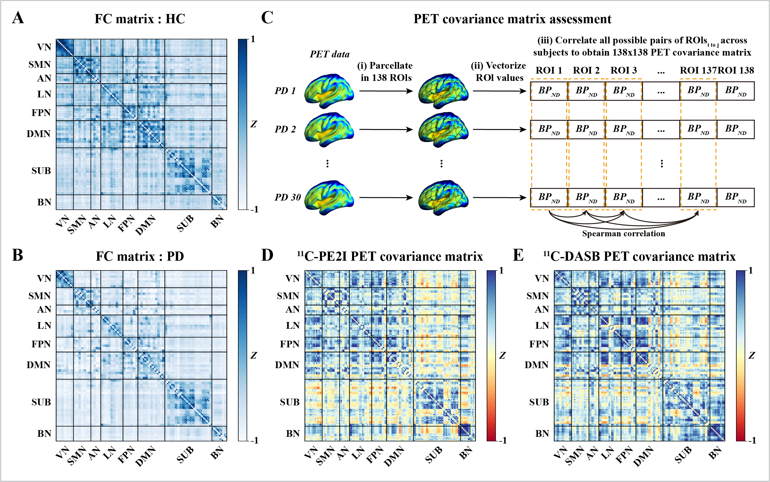
Method: We generated FC matrices and assessed the covariance of 11C-PE2I and 11C-DASB PET uptake across 138 ROIs using the AAL3 atlas in 30 PD patients at baseline, with a 19-month follow-up for 15 of these patients. Linear regression was used to assess the association between FC and PET covariance. The same analysis methods were used longitudinally, substituting baseline PET uptake with change between baseline and follow-up. Finally, FC-PET association (b) maps for each PD patient and tracer were generated, and Spearman’s correlation was used to identify brain regions where these coefficients correlated with PD severity.
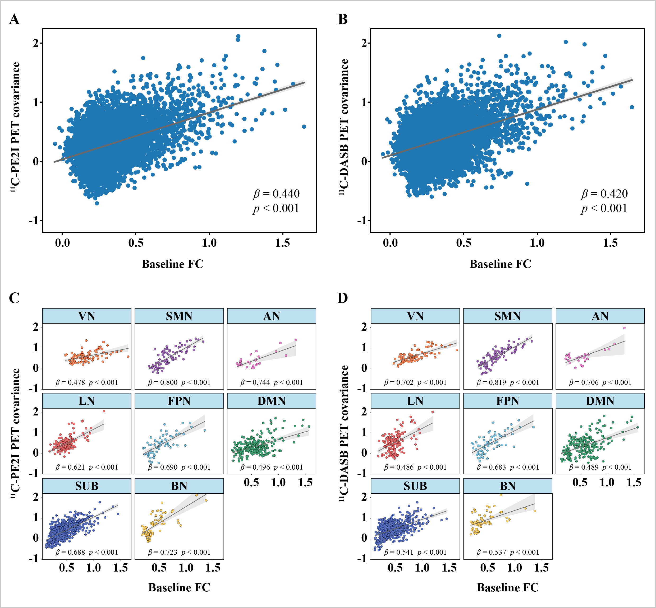
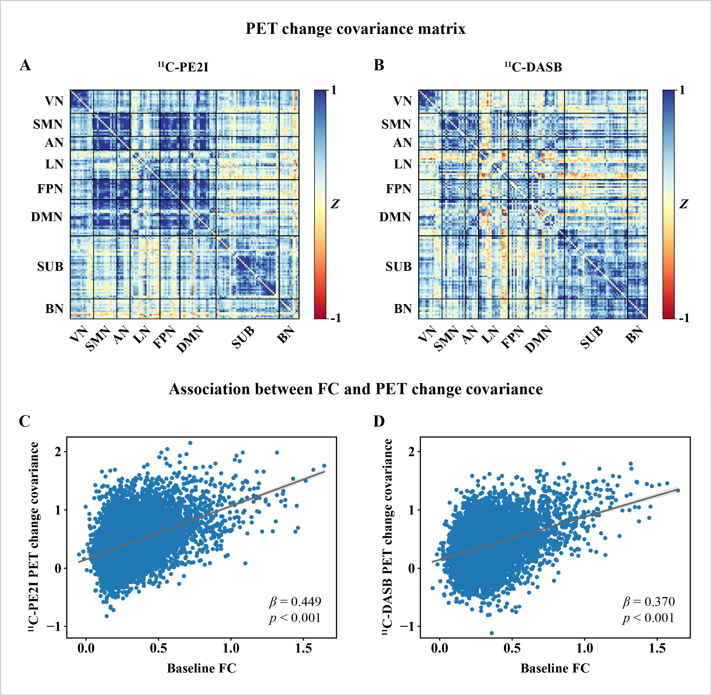
Results: At baseline, linear regression showed that FC was positively related to both 11C-PE2I PET covariance (beta=0.440, p<0.001) and 11C-DASB PET covariance (beta=0.420, p<0.001). Longitudinally, positive correlations were found between baseline FC and both 11C-PE2I PET change covariance (beta=0.449, p<0.001) and 11C-DASB PET change covariance (beta=0.370, p<0.001). For both tracers, absolute PET uptake across seed ROIs was positively associated with correspondent regression-derived FC-PET beta-weights, representing the relationship between PET uptake in target ROIs and their functional connectivity to the seed. This association between target FC and PET uptake was correlated with PD motor and non-motor severity across different brain regions that was dependent on the neurotransmitter system evaluated.
Conclusion: In PD patients, brain regions with high FC exhibit similar dopamine and serotonin levels, as well as parallel longitudinal changes in these neurotransmitter levels. Our findings indicate a significant correspondence between FC patterns and the spatial distribution of dopamine and serotonin.
Group-average matrices at baseline.
FC and PET covariance association.
FC and PET change covariance association.
To cite this abstract in AMA style:
W. Li, N. Lao-Kaim, R. Li, A. Martín-Bastida, A. Roussakis, G. Searle, N. Valle-Guzman, V. Dayal, D. Athauda, Z. Kefalopoulou, P. Mahlknecht, A. Church, K. Peall, H. Widner, G. Paul, T. Foltynie, R. Barker, P. Piccini. Mapping the distribution of neurotransmitters to resting-state functional connectivity in patients with Parkinson’s disease [abstract]. Mov Disord. 2024; 39 (suppl 1). https://www.mdsabstracts.org/abstract/mapping-the-distribution-of-neurotransmitters-to-resting-state-functional-connectivity-in-patients-with-parkinsons-disease/. Accessed December 12, 2025.« Back to 2024 International Congress
MDS Abstracts - https://www.mdsabstracts.org/abstract/mapping-the-distribution-of-neurotransmitters-to-resting-state-functional-connectivity-in-patients-with-parkinsons-disease/


