Category: Parkinson's Disease: Neuroimaging
Objective: To validate the previously characterized resting-state fMRI (rs-fMRI) Parkinson’s disease (PD)-related pattern, termed fPDRP, in a large multi-site PD imaging database, and to assess changes in expression occurring with disease progression.
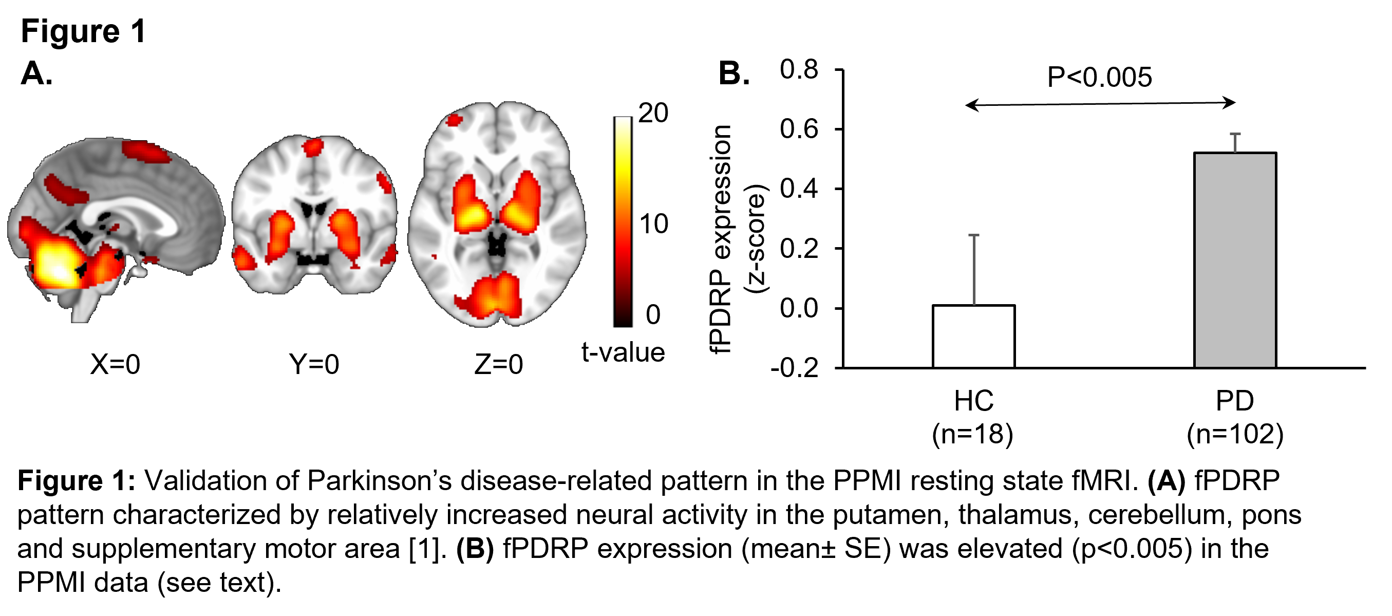
Background: We recently identified an fPDRP network (Figure 1A) using a new computational method based on independent component analysis (ICA) and bootstrap resampling [1], which we subsequently validated in an independent rs-fMRI dataset [2]. Significant elevations in fPDRP expression were observed in PD vs. healthy control (HC) subjects in both samples [1-2]. It is unclear, however, whether expression levels for this pattern change over time in longitudinal rs-fMRI scans from PD patients.
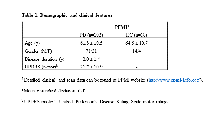
Method: We analyzed rs-fMRI scans from 102 PD and 18 HC (Table 1) who were studied as part of Parkinson’s Progression Markers Initiative (PPMI; https://www.ppmi-info.org/). All patients and control subjects were scanned at baseline; 43 PD were rescanned at 1-year and 35 at 2-years. Subject spatial maps were estimated for each scan using dual regression [1-3]. Individual fPDRP expression values were computed and z-scored with respect to the HC distribution. fPDRP expression was computed at patient and time point and changes from baseline were evaluated at 12- and 24-months using paired Student’s t-tests.
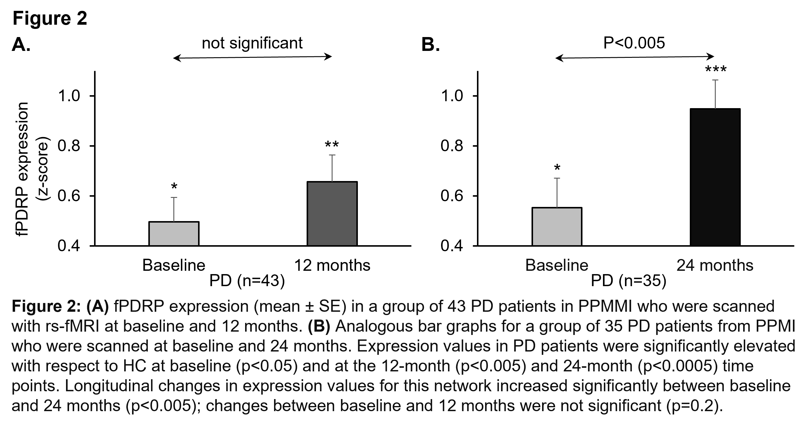
Results: fPDRP expression was elevated in baseline rs-fMRI scans from PD patients in PPMI relative to HC subjects (n=18) from the same population (p< 0.005, Student’s t-test; Figure 1B). fPDRP expression levels increased on follow-up (Figure 2A,B) with greater deviations from HC at each successive time point (12 months: p<0.005; 24 months: p<0.0005). Compared to baseline, longitudinal increases in fPDRP expression were significant at 24 months (p<0.005) but not at 12 months.
Conclusion: In the PPMI PD cohort, fPDRP exhibited significant abnormal elevations and changes in network scores over a 2-year period. The result suggests that fPDRP may be useful as a biomarker in multi-site rs-fMRI data from PD patients.
References: References:
1. Vo, A. (2017), ‘Parkinson’s disease-related network topographies characterized with resting state functional MRI’, Hum Brain Mapp, vol. 38, no. 2, pp. 617–630.
2. Rommal. A, (2021), ‘Parkinson’s disease-related pattern (PDRP) identified using resting-state functional MRI: Validation study’, Neuroimage: Reports, vol. 1, no. 3, pp. 100026
3. Schindlbeck, K. (2021), ‘Cognition-related functional topographies in Parkinson’s disease: Localized loss of the ventral default mode network’. Cerebral Cortex, vol. 31, no. 11, pp. 5139-5150.
To cite this abstract in AMA style:
A. Vo, N. Nguyen, D. Eidelberg. Longitudinal study of Parkinson’s disease-related pattern with resting state functional MRI [abstract]. Mov Disord. 2022; 37 (suppl 2). https://www.mdsabstracts.org/abstract/longitudinal-study-of-parkinsons-disease-related-pattern-with-resting-state-functional-mri/. Accessed February 23, 2026.« Back to 2022 International Congress
MDS Abstracts - https://www.mdsabstracts.org/abstract/longitudinal-study-of-parkinsons-disease-related-pattern-with-resting-state-functional-mri/


